 Back
BackMicroscopy: Observing Microorganisms Through a Microscope
Study Guide - Smart Notes
 Tailored notes based on your materials, expanded with key definitions, examples, and context.
Microscopy: Observing Microorganisms Through a Microscope
Introduction to Microscopy
Microscopy is essential in microbiology for visualizing organisms too small to be seen with the naked eye. Various types of microscopes and staining techniques allow scientists to study the structure, function, and classification of microorganisms.
Units of Measurement in Microbiology
Micrometers (μm) and nanometers (nm) are the standard units for measuring microorganisms.
1 μm = meters
1 nm = meters
1000 nm = 1 μm
Microorganisms typically range from 1 μm to several micrometers in size.
Types of Microscopes
Simple Microscope
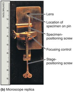
A simple microscope uses a single lens for magnification, similar to a magnifying glass but with higher quality optics. Anton van Leeuwenhoek's early observations were made with such an instrument.
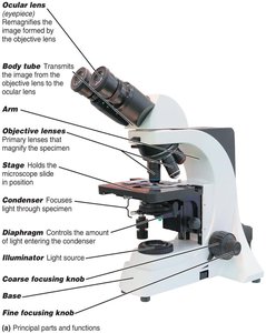
Compound Light Microscope
The compound light microscope uses multiple lenses to achieve higher magnification and resolution. It is the most common microscope in microbiology labs.
Ocular lens (eyepiece): Remagnifies the image from the objective lens.
Objective lenses: Primary lenses that magnify the specimen (commonly 4x, 10x, 40x, 100x).
Stage: Holds the microscope slide.
Condenser: Focuses light through the specimen.
Diaphragm: Controls the amount of light entering the condenser.
Illuminator: Light source.
Coarse and fine focusing knobs: Adjust the focus.
Path of Light in a Compound Microscope
Light passes from the illuminator through the condenser, specimen, objective lens, body tube, and ocular lens to the observer's eye. The image is magnified at each stage.

Total Magnification and Resolution
Total Magnification: Product of the magnification of the objective lens and the ocular lens.
Example: 40x objective × 10x ocular = 400x total magnification.
Resolution (resolving power): The ability to distinguish two points as separate. Higher resolution allows for clearer, more detailed images.
Shorter wavelengths of light provide greater resolution.
The limit of resolution for a compound light microscope is about 0.2 μm.
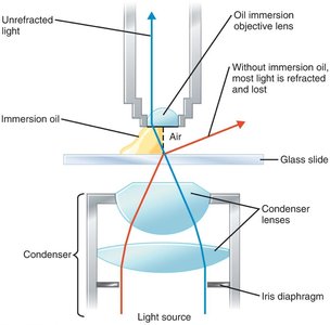
Refractive Index and Immersion Oil
The refractive index measures how much a medium bends light. Immersion oil is used with high-power objectives to reduce light refraction and increase resolution.
Types of Light Microscopy
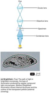
Brightfield Microscopy
Brightfield microscopy is the standard form of light microscopy, where dark objects are visible against a bright background. It is best for stained specimens.
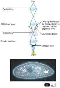
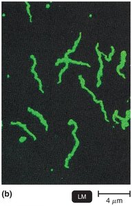
Darkfield Microscopy
Darkfield microscopy uses an opaque disk to block direct light, so only light reflected by the specimen enters the objective lens. This technique is useful for viewing live, unstained microorganisms, such as Treponema pallidum.
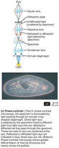
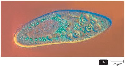
Phase-Contrast Microscopy
Phase-contrast microscopy enhances contrast in transparent specimens without staining. It combines direct and diffracted light rays to visualize internal structures in living cells.
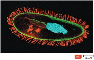
Differential Interference Contrast (DIC) Microscopy
DIC microscopy uses two beams of light and prisms to produce high-contrast, brightly colored, three-dimensional images of specimens.
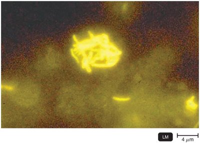
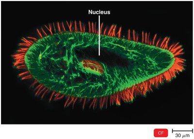
Fluorescence Microscopy
Fluorescence microscopy uses ultraviolet (UV) light to excite fluorescent dyes (fluorochromes) that emit visible light. It is widely used for detecting specific microbes using fluorescent antibodies (immunofluorescence).
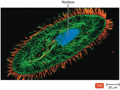
Confocal Microscopy
Confocal microscopy uses lasers and fluorochromes to scan specimens in layers, producing sharp, two-dimensional images that can be reconstructed into three-dimensional models.
Two-Photon Microscopy
Two-photon microscopy uses two photons of long-wavelength light to excite fluorochromes, allowing imaging of living cells up to 1 mm deep and tracking cell activity in real time.
Super-Resolution Light Microscopy
Super-resolution microscopy uses advanced laser techniques to surpass the diffraction limit of light, enabling visualization of structures at the nanometer scale and tracking single molecules in living cells.
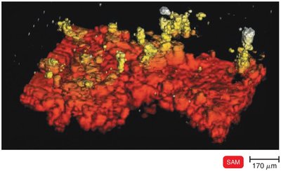
Scanning Acoustic Microscopy (SAM)
SAM uses sound waves to image specimens, particularly useful for studying cells attached to surfaces, such as biofilms and cancer cells. It has a resolution of about 1 μm.
Electron Microscopy
Electron microscopes use electron beams instead of light, providing much higher resolution and magnification. They are essential for visualizing viruses and internal cell structures.
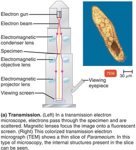
Transmission Electron Microscopy (TEM)
Electrons pass through ultrathin sections of a specimen.
Magnification: 10,000–10,000,000x; Resolution: 0.2 nm.
Used for detailed internal structure imaging.
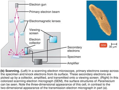
Scanning Electron Microscopy (SEM)
Electron beam scans the surface of a specimen.
Secondary electrons emitted from the surface are collected to form a 3D image.
Magnification: 1,000–500,000x; Resolution: 0.5 nm.
Used for detailed surface imaging.
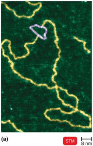
Scanned-Probe Microscopy
Scanned-probe microscopes use physical probes to scan specimen surfaces, providing atomic or near-atomic resolution without specimen modification.
Scanning Tunneling Microscopy (STM): Uses a tungsten probe to scan surfaces, resolving features as small as atoms.
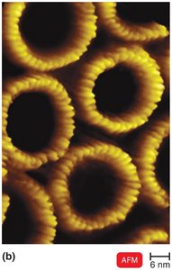
Atomic Force Microscopy (AFM): Uses a metal-and-diamond probe to produce 3D images at near-atomic detail.
Staining Techniques in Microbiology
Preparing Smears for Staining
Staining: Coloring microorganisms with dyes to emphasize structures.
Smear: Thin film of microorganisms spread on a slide.
Fixation: Attaches and kills microorganisms, preserving their structure. Methods include heat or chemical fixation (e.g., methanol).
Types of Dyes
Basic dyes: Chromophore is a cation (e.g., crystal violet, methylene blue, safranin). Adheres to negatively charged bacterial cells.
Acidic dyes: Chromophore is an anion (e.g., eosin, acid fuchsin, nigrosin). Used for negative staining (stains background, not cells).
Simple Stains
Simple stains use a single basic dye to highlight the entire microorganism, making cell shapes and structures visible. A mordant may be used to enhance staining.

Differential Stains
Differential stains distinguish between different types of bacteria. The most common are the Gram stain and the acid-fast stain.
Gram Stain
Classifies bacteria as gram-positive (thick peptidoglycan, purple) or gram-negative (thin peptidoglycan, outer membrane, pink/red).
Steps:
Primary stain: Crystal violet (both types purple)
Mordant: Gram's iodine (both types purple)
Decolorizer: Alcohol/acetone (gram-positive purple, gram-negative colorless)
Counterstain: Safranin (gram-positive purple, gram-negative pink/red)
Acid-Fast Stain
Identifies bacteria with waxy cell walls (e.g., Mycobacterium, Nocardia).
Steps:
Primary stain: Carbolfuchsin (all cells red)
Decolorizer: Acid-alcohol (acid-fast red, non–acid-fast colorless)
Counterstain: Methylene blue (acid-fast red, non–acid-fast blue)
Special Stains
Special stains are used to highlight specific structures:
Capsule stain: Visualizes the gelatinous capsule surrounding some bacteria.
Endospore stain: Detects highly resistant spores within bacteria.
Flagella stain: Visualizes bacterial flagella for motility studies.
Summary Table: Types of Microscopy and Their Applications
Microscopy Type | Principle | Best Use |
|---|---|---|
Brightfield | Light passes through specimen | Stained cells, general morphology |
Darkfield | Only reflected light enters objective | Live, unstained cells; spirochetes |
Phase-Contrast | Combines direct and diffracted light | Internal structures of live cells |
DIC | Two beams, prisms for 3D effect | 3D, high-contrast images |
Fluorescence | UV light excites fluorochromes | Specific detection with antibodies |
Confocal | Laser scans layers | 3D reconstructions |
Two-Photon | Two photons excite dye | Deep tissue, live cell imaging |
Super-Resolution | Advanced lasers, computation | Single-molecule, nanometer scale |
SAM | Sound waves | Surface-attached cells, biofilms |
TEM | Electrons through specimen | Internal ultrastructure |
SEM | Electrons scan surface | 3D surface structure |
STM/AFM | Physical probe scans surface | Atomic/molecular detail |
