 Back
BackMicroscopy: Observing Microorganisms Through a Microscope
Study Guide - Smart Notes
 Tailored notes based on your materials, expanded with key definitions, examples, and context.
Microscopy: Observing Microorganisms Through a Microscope
Units of Measurement in Microbiology
Microorganisms are measured using the metric system, primarily in micrometers (µm) and nanometers (nm). Understanding these units is essential for interpreting microscopic observations.
Micrometer (µm): 1 µm = 10-6 meters
Nanometer (nm): 1 nm = 10-9 meters
1 µm = 1000 nm
Example: Most bacteria are 1–10 µm in length, while viruses range from 20–300 nm.
Types of Microscopes
Microscopes are essential tools for visualizing microorganisms. The two main categories are light microscopes and electron microscopes.
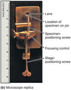
Simple Microscope
A simple microscope uses a single lens for magnification, similar to a magnifying glass but with higher quality optics.
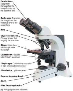
Compound Light Microscope
The compound light microscope uses multiple lenses to achieve higher magnification and resolution. It is the most common instrument in microbiology labs.
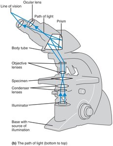
Path of Light in a Compound Microscope
Light passes from the illuminator through the condenser, specimen, objective lens, body tube, and finally the ocular lens to the observer's eye. The image is magnified at each stage.
Total Magnification and Resolution
Total Magnification is calculated by multiplying the magnification of the objective lens by that of the ocular lens:
Resolution (resolving power) is the ability to distinguish two points as separate entities. The limit of resolution for a compound light microscope is about 0.2 µm. Shorter wavelengths of light provide greater resolution.
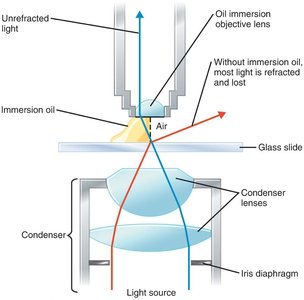
Refractive Index and Immersion Oil
The refractive index is a measure of how much a substance bends light. Immersion oil is used with high-power objectives to reduce light refraction and increase resolution by matching the refractive index of glass.
Types of Light Microscopy
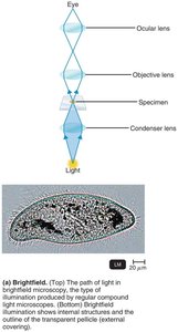
Brightfield Microscopy
Brightfield microscopy is the standard form of light microscopy. It produces a dark image on a bright background and is suitable for stained specimens.
Light passes directly through the specimen.
Unstained cells may be difficult to see due to low contrast.
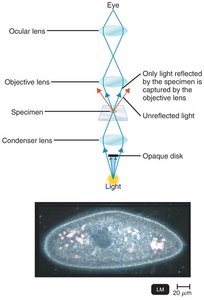
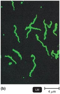
Darkfield Microscopy
Darkfield microscopy enhances the contrast in unstained samples. Only light reflected by the specimen enters the objective lens, making the specimen appear bright against a dark background.
Useful for observing live, unstained microorganisms (e.g., Treponema pallidum).
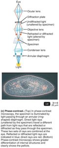
Phase-Contrast Microscopy
Phase-contrast microscopy allows for the detailed examination of living cells and their internal structures without staining. It uses differences in refractive index to enhance contrast.
Combines direct and diffracted light rays to form an image.
Ideal for observing cellular processes in real time.
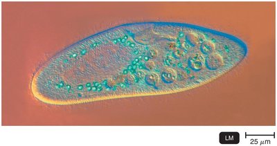
Differential Interference Contrast (DIC) Microscopy
DIC microscopy is similar to phase-contrast but uses two beams of light and prisms to produce high-contrast, brightly colored, and three-dimensional images of specimens.
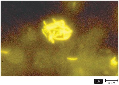
Fluorescence Microscopy
Fluorescence microscopy uses ultraviolet (UV) light to excite fluorescent dyes (fluorochromes) that emit visible light. It is widely used for detecting specific microorganisms and cellular components.
Cells may be stained with fluorochromes if they do not naturally fluoresce.
Immunofluorescence uses antibodies tagged with fluorochromes for specific detection of pathogens.
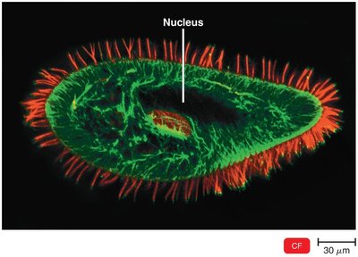
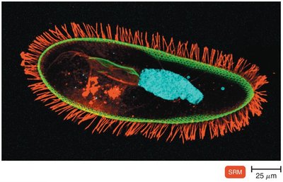
Confocal Microscopy
Confocal microscopy uses laser light to scan specimens stained with fluorochromes, producing sharp, two-dimensional images at various depths. Computer reconstruction allows for three-dimensional visualization.
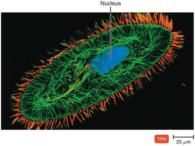
Two-Photon Microscopy
Two-photon microscopy uses long-wavelength (red) light to excite fluorochromes, allowing imaging of living cells up to 1 mm deep and tracking cellular activity in real time.
Super-Resolution Light Microscopy
Super-resolution microscopy surpasses the diffraction limit of light, enabling visualization of structures at the nanometer scale using advanced laser techniques and computational reconstruction.
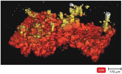
Scanning Acoustic Microscopy (SAM)
SAM uses sound waves reflected from a specimen to generate images. It is useful for studying cells attached to surfaces, such as biofilms, with a resolution of about 1 µm.
Electron Microscopy
Electron microscopes use electron beams instead of light, providing much higher resolution and magnification. They are essential for visualizing viruses and internal cellular structures.
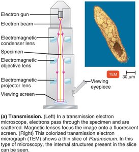
Transmission Electron Microscopy (TEM)
TEM passes electrons through ultrathin sections of specimens, revealing internal structures at very high resolution (up to 0.2 nm). Specimens must be fixed, dehydrated, and sectioned, which kills the cells.
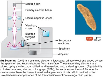
Scanning Electron Microscopy (SEM)
SEM scans the surface of a specimen with a focused electron beam, producing detailed three-dimensional images of surface structures. The resolution is about 0.5 nm.
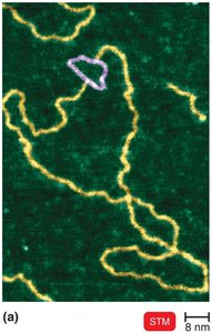
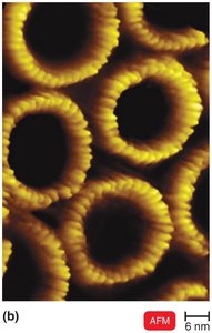
Scanned-Probe Microscopy
Scanned-probe microscopes use physical probes to scan specimen surfaces, allowing for atomic and molecular resolution without specimen modification.
Scanning Tunneling Microscopy (STM): Uses a tungsten probe to scan surfaces, resolving features as small as atoms.
Atomic Force Microscopy (AFM): Uses a metal-and-diamond probe to produce three-dimensional images at near-atomic detail.
Staining and Preparing Microbial Specimens
Staining Techniques
Staining enhances contrast in microscopic images by coloring microorganisms or their background. Fixation (by heat or chemicals) attaches and preserves cells on the slide.
Basic dyes: Chromophore is a cation (e.g., crystal violet, methylene blue, safranin).
Acidic dyes: Chromophore is an anion (e.g., eosin, acid fuchsin, nigrosin).
Negative staining: Stains the background, not the cell, using acidic dyes.
Simple Staining
Simple stains use a single basic dye to highlight the entire microorganism, making cell shapes and structures visible. A mordant may be used to intensify the stain.

Differential Staining
Differential stains distinguish between different types of bacteria. The two most common are the Gram stain and the acid-fast stain.
Gram Stain
The Gram stain classifies bacteria as gram-positive (thick peptidoglycan, purple) or gram-negative (thin peptidoglycan, outer membrane, pink/red). It is a critical diagnostic tool in microbiology.
Step | Gram-Positive | Gram-Negative |
|---|---|---|
Primary Stain: Crystal Violet | Purple | Purple |
Mordant: Gram’s Iodine | Purple | Purple |
Decolorizing Agent: Alcohol/Acetone | Purple | Colorless |
Counterstain: Safranin | Purple | Pink/Red |
Acid-Fast Stain
The acid-fast stain identifies bacteria with waxy cell walls (e.g., Mycobacterium). Acid-fast cells retain the primary stain (red) after acid-alcohol decolorization, while non–acid-fast cells take up the counterstain (blue).
Step | Acid-Fast | Non–Acid-Fast |
|---|---|---|
Primary Stain: Carbolfuchsin | Red | Red |
Decolorizing Agent: Acid-Alcohol | Red | Colorless |
Counterstain: Methylene Blue | Red | Blue |
Special Stains
Special stains are used to highlight specific structures:
Capsule stain: Visualizes the gelatinous capsule surrounding some bacteria.
Endospore stain: Detects resistant, dormant structures within bacteria.
Flagella stain: Reveals the presence and arrangement of flagella.
Summary Table: Types of Microscopy and Their Applications
Microscopy Type | Principle | Application |
|---|---|---|
Brightfield | Light passes through specimen | Stained cells, general morphology |
Darkfield | Only reflected light enters lens | Live, unstained cells |
Phase-Contrast | Enhances differences in refractive index | Internal structures of live cells |
DIC | Two beams, prisms for 3D effect | 3D images, color contrast |
Fluorescence | UV light excites fluorochromes | Specific detection, immunofluorescence |
Confocal | Laser scans stained specimen | 3D reconstruction, biofilms |
Two-Photon | Two photons excite dye | Deep tissue imaging |
Super-Resolution | Advanced lasers, computation | Single-molecule tracking |
SAM | Sound waves | Surface-attached cells, biofilms |
TEM | Electrons through specimen | Internal ultrastructure |
SEM | Electrons scan surface | Surface morphology, 3D images |
STM/AFM | Physical probe scans surface | Atomic/molecular detail |
