 Back
BackMicroscopy: Principles, Types, and Applications in Microbiology
Study Guide - Smart Notes
 Tailored notes based on your materials, expanded with key definitions, examples, and context.
Microscopy in Microbiology
Units of Measurement
Microorganisms are measured using the metric system, primarily in micrometers (μm) and nanometers (nm). Understanding these units is essential for interpreting microscopic images and comparing microbial sizes.
Micrometer (μm): 1 μm = 10-6 meters
Nanometer (nm): 1 nm = 10-9 meters
Conversion: 1 μm = 1000 nm
Types of Microscopes
Microscopes are essential tools in microbiology, allowing visualization of organisms too small for the naked eye. There are several types, each with unique features and applications.
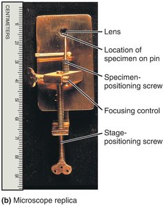
Simple Microscope: Contains a single lens, similar to a magnifying glass but with higher magnification.
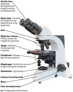
Compound Light Microscope: Uses multiple lenses to magnify specimens and is the most common in laboratories.
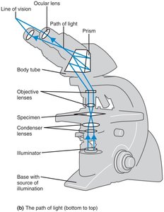
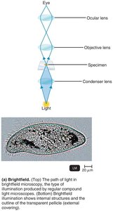
Path of Light in a Compound Microscope
The compound microscope uses a series of lenses to direct light through the specimen, magnifying the image for observation.
Light originates from the illuminator and passes through the condenser lenses.
It then travels through the specimen, objective lenses, and body tube.
Finally, the light passes through the ocular lens (eyepiece) to the observer's eye.
Total Magnification and Resolution
Total magnification is calculated by multiplying the magnification of the objective lens by that of the ocular lens. Resolution (resolving power) is the ability to distinguish two points as separate entities.
Total Magnification Formula:
Resolution: Shorter wavelengths of light provide greater resolution. The limit for compound light microscopes is about 0.2 μm.
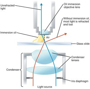
Refractive Index and Immersion Oil
The refractive index measures the light-bending ability of a medium. Immersion oil is used with high-power objective lenses to prevent light from refracting away from the lens, thus improving resolution.
Immersion oil has a refractive index similar to glass, keeping the light path straight.
Without oil, light is refracted and lost, reducing image clarity.
Types of Light Microscopy
Brightfield Microscopy
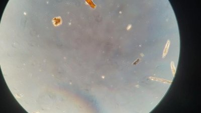
Brightfield microscopy is the standard method, where dark objects are visible against a bright background. It is suitable for stained specimens but may lack contrast for unstained cells.
Shows internal structures and cell outlines.
May require staining for better visualization.
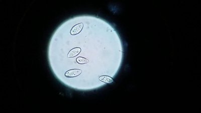
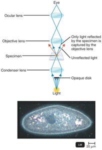
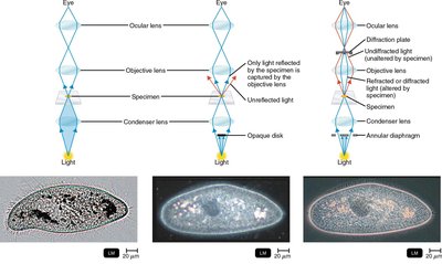
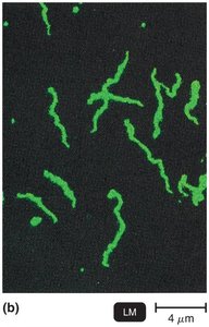
Darkfield Microscopy
Darkfield microscopy uses a special condenser with an opaque disk, making light objects visible against a dark background. Only light reflected by the specimen enters the objective lens.
Useful for viewing live, unstained microorganisms.
Can visualize slender bacteria such as Treponema pallidum.
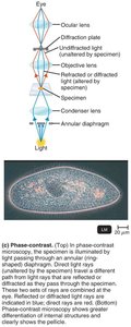
Phase-Contrast Microscopy
Phase-contrast microscopy allows detailed examination of living organisms and internal cell structures without staining. It combines direct and diffracted light rays to enhance contrast.
Shows greater differentiation of internal structures.
Ideal for observing live cells.
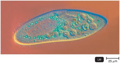
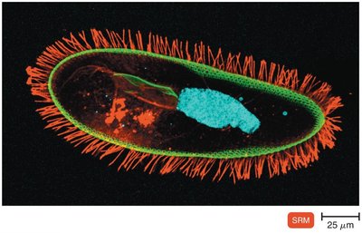
Differential Interference Contrast (DIC) Microscopy
DIC microscopy is similar to phase-contrast but uses two light beams and prisms to produce high-contrast, brightly colored, and three-dimensional images.
Provides enhanced contrast and color.
Images appear three-dimensional.
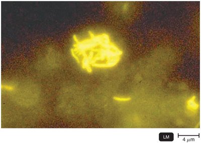
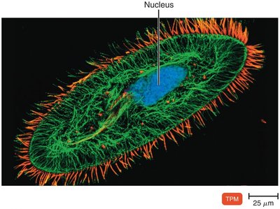
Fluorescence Microscopy
Fluorescence microscopy uses UV light to excite fluorescent substances, which emit visible light. Cells may be stained with fluorochromes for visualization.
Allows rapid and specific detection of pathogens.
Fluorescent-antibody technique (immunofluorescence) is used for diagnostic purposes.
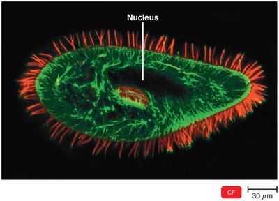
Confocal Microscopy
Confocal microscopy uses fluorochrome dyes and laser light to produce clear two-dimensional images of a single plane. Multiple planes can be combined to create three-dimensional images.
Provides high-resolution, three-dimensional reconstructions.
Useful for studying cell structures and biofilms.
Two-Photon Microscopy
Two-photon microscopy uses two photons of long-wavelength light to excite dyes, allowing the study of living cells up to 1 mm deep and tracking cell activity in real time.
Enables deep tissue imaging.
Useful for observing live cell dynamics.
Super-Resolution Light Microscopy
Super-resolution microscopy uses two laser beams to achieve nanometer-scale resolution, allowing single-molecule tracking and detailed study of protein movements in cells.
Provides resolution beyond the diffraction limit of light.
Used for advanced cellular and molecular studies.
Scanning Acoustic Microscopy
Scanning acoustic microscopy measures sound waves reflected from a specimen, useful for studying cells attached to surfaces such as biofilms, cancer cells, and arterial plaque.
Resolution is about 1 μm.
Non-invasive technique for surface studies.

Electron Microscopy
Principles of Electron Microscopy
Electron microscopes use electron beams instead of light, providing much greater resolution due to the shorter wavelength of electrons. They are essential for visualizing viruses and internal cellular structures.
Images are black and white, often colorized digitally.
Use electromagnetic lenses to focus electron beams.
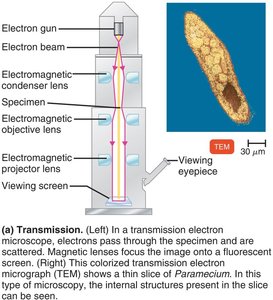
Transmission Electron Microscopy (TEM)
TEM passes electrons through ultrathin sections of a specimen, producing highly detailed images of internal structures.
Magnification: 10,000–10,000,000x
Resolution: 0.2 nm
Specimen preparation involves fixation, dehydration, and slicing.
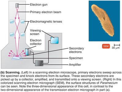
Scanning Electron Microscopy (SEM)
SEM scans the surface of specimens with an electron beam, providing three-dimensional images of surface structures.
Magnification: 1,000–500,000x
Resolution: 0.5 nm
Ideal for studying surface morphology.
Scanned-Probe Microscopy
Principles and Types
Scanned-probe microscopes use probes to examine specimen surfaces with electric current, enabling atomic and molecular mapping without specimen modification.
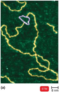
Scanning Tunneling Microscopy (STM): Uses a tungsten probe to scan surfaces, resolving features as small as atoms.
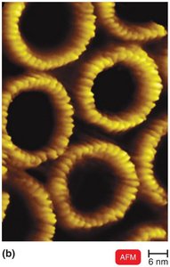
Atomic Force Microscopy (AFM): Uses a metal-and-diamond probe to produce three-dimensional images at near atomic detail.
Staining Techniques in Microbiology
Preparing Smears for Staining
Staining is used to color microorganisms, emphasizing certain structures. Smears are thin films of material containing microorganisms spread over a slide and fixed before staining.
Fixation: Attaches and kills microorganisms, preserving structures.
Methods: Heat fixation or chemical fixation (methanol).
Acidic vs. Basic Dyes
Dyes consist of a colored ion (chromophore). In basic dyes, the chromophore is a cation; in acidic dyes, it is an anion. Bacterial cells are negatively charged, so basic dyes adhere to them.
Basic dyes: Crystal violet, methylene blue, safranin
Acidic dyes: Eosin, acid fuchsin, nigrosin
Negative staining: Stains the background using acidic dyes
Simple Staining
Simple stains use a single basic dye to highlight the entire microorganism, making cell shapes and structures visible. A mordant may be used to enhance staining.
Examples: Methylene blue, carbolfuchsin, crystal violet, safranin
Differential Stains
Differential stains distinguish between types of bacteria. The most common are the Gram stain and acid-fast stain.
Gram Stain: Classifies bacteria as gram-positive (purple, thick peptidoglycan) or gram-negative (pink/red, thin peptidoglycan and outer membrane).
Acid-Fast Stain: Identifies bacteria with waxy cell walls, such as Mycobacterium and Nocardia.
Special Stains
Special stains are used to distinguish specific parts of microorganisms, such as capsules, endospores, and flagella.
Capsule stain: Highlights the capsule surrounding some bacteria.
Endospore stain: Identifies endospores within bacterial cells.
Flagella stain: Visualizes flagella for motility studies.
Summary Table: (For review, see Table 3.2 in the textbook.)
Additional info: These notes cover the essential principles, types, and applications of microscopy in microbiology, including staining techniques and the interpretation of microscopic images. Images included are directly relevant to the described microscopy methods and staining procedures.
