 Back
BackMicroscopy, Staining, and Cell Structure in Microbiology
Study Guide - Smart Notes
 Tailored notes based on your materials, expanded with key definitions, examples, and context.
Observing Microorganisms through a Microscope
Differential Stains
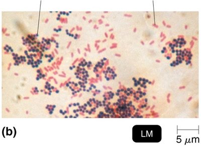
Differential stains are essential tools in microbiology for classifying bacteria based on their cell wall structure. The most common differential stain is the Gram stain, which separates bacteria into gram-positive and gram-negative groups.
Gram-positive bacteria: Have thick peptidoglycan cell walls.
Gram-negative bacteria: Have thin peptidoglycan cell walls and an outer layer of lipopolysaccharides.
Gram Staining Procedure
The Gram stain involves a series of steps that result in different coloration of bacterial cells, depending on their cell wall structure.
Application of crystal violet (primary stain)
Application of iodine (mordant)
Alcohol wash (decolorization)
Application of safranin (counterstain)
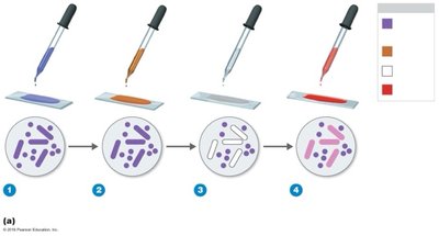
Gram-positive cells: Remain purple throughout the process.
Gram-negative cells: Become colorless after alcohol wash, then turn red after safranin is applied.
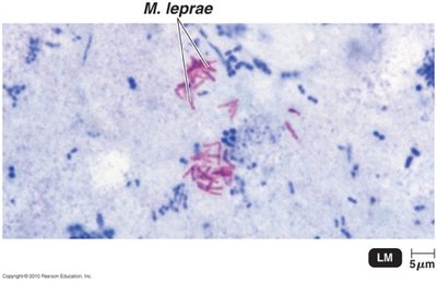
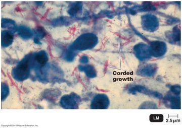
Acid-Fast Stain
The acid-fast stain is used to identify bacteria with waxy cell walls, such as Mycobacterium and Nocardia. These bacteria retain the primary stain even after decolorization with acid-alcohol.
Acid-fast bacteria: Stain red with carbolfuchsin.
Non–acid-fast bacteria: Stain blue with methylene blue after decolorization.
Special Stains
Special stains are used to highlight specific structures within microorganisms, such as capsules, endospores, and flagella.
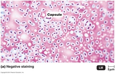
Capsule stain: Reveals the presence of a capsule, which is a protective layer outside the cell wall.
Endospore stain: Identifies resistant, dormant structures within cells.
Flagella stain: Makes flagella visible by thickening them with mordant and carbolfuchsin.
Cell Structure and Function
Comparing Prokaryotic and Eukaryotic Cells
Prokaryotic and eukaryotic cells differ in their genetic material, organelles, and cell wall composition.
Prokaryotes: One circular chromosome, no membrane-bound organelles, peptidoglycan cell walls (bacteria), divide by binary fission.
Eukaryotes: Paired chromosomes in a nuclear membrane, organelles present, polysaccharide cell walls (when present), divide by mitosis.
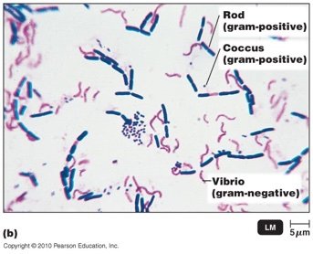
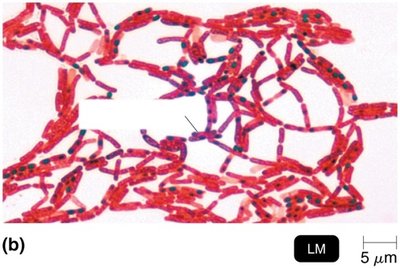
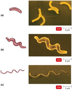
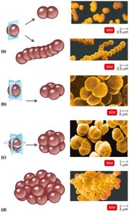
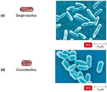
The Size, Shape, and Arrangement of Bacterial Cells
Bacteria exhibit a variety of shapes and arrangements, which are important for identification and classification.
Size: 0.2 to 2.0 μm diameter × 2 to 8 μm length
Shapes: Bacillus (rod-shaped), Coccus (spherical), Spiral (vibrio, spirillum, spirochete), star-shaped, rectangular
Arrangements: Diplococci, Streptococci, Tetrad, Sarcinae, Staphylococci
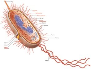
The Structure of a Prokaryotic Cell
Prokaryotic cells have a simple structure, lacking membrane-bound organelles. Key components include the cell wall, plasma membrane, cytoplasm, nucleoid, plasmids, ribosomes, and external structures such as flagella, fimbriae, and pili.

The Cell Wall
The cell wall provides structural support and protection. In bacteria, it is composed of peptidoglycan, a polymer of N-acetylglucosamine (NAG) and N-acetylmuramic acid (NAM) linked by polypeptides.
Gram-positive cell wall: Thick peptidoglycan, teichoic acids, disrupted by lysozyme, sensitive to penicillin.
Gram-negative cell wall: Thin peptidoglycan, outer membrane, periplasmic space, endotoxin, sensitive to tetracycline.
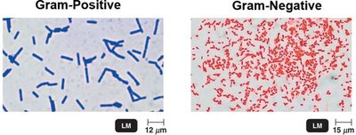
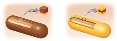
Table: Comparative Characteristics of Gram-Positive and Gram-Negative Bacteria
Feature | Gram-Positive | Gram-Negative |
|---|---|---|
Peptidoglycan | Thick | Thin |
Teichoic acids | Present | Absent |
Outer membrane | Absent | Present |
Basal body rings | 2 | 4 |
Lysozyme sensitivity | Yes | No |
Penicillin sensitivity | Yes | No |
Endotoxin | No | Yes |
Atypical Cell Walls
Some bacteria have atypical cell walls:
Acid-fast cell walls: Like gram-positive, but with waxy lipid (mycolic acid) bound to peptidoglycan. Found in Mycobacterium and Nocardia.
Mycoplasmas: Lack cell walls, have sterols in plasma membrane.
Archaea: May lack cell walls or have walls of pseudomurein (lack NAM and D-amino acids).
Damage to the Cell Wall
Cell wall-targeting agents are important in antimicrobial therapy.
Lysozyme: Hydrolyzes bonds in peptidoglycan.
Penicillin: Inhibits peptide bridges in peptidoglycan.
Protoplast: Wall-less gram-positive cell.
Spheroplast: Wall-less gram-negative cell.
L forms: Wall-less cells that swell into irregular shapes.
Glycocalyx
The glycocalyx is an external, viscous, gelatinous layer made of polysaccharide. It contributes to virulence by preventing phagocytosis and helping form biofilms.
Capsule: Neatly organized and firmly attached.
Slime layer: Unorganized and loose.
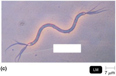
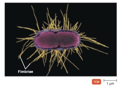
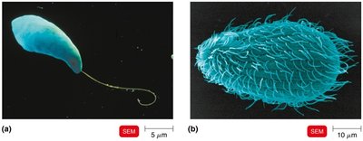
Flagella, Fimbriae, and Pili
These structures are important for motility, attachment, and genetic exchange.
Flagella: Filamentous appendages for locomotion, made of flagellin.
Fimbriae: Hairlike appendages for attachment.
Pili: Involved in motility and DNA transfer (conjugation pili).
Eukaryotic Flagella and Cilia
Eukaryotic cells may possess flagella or cilia for movement. These structures are more complex than their prokaryotic counterparts.
Flagella: Long, whip-like structures for movement.
Cilia: Short, numerous appendages for movement or moving substances along the cell surface.
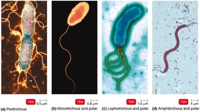
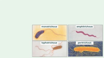
Flagella Arrangement and Motility
Bacterial flagella can be arranged in different patterns, affecting their motility.
Monotrichous: Single flagellum at one end.
Lophotrichous: Cluster of flagella at one end.
Amphitrichous: Flagella at both ends.
Peritrichous: Flagella distributed over the entire cell.
Check Your Understanding
Why are drugs that target cell wall synthesis useful? They selectively inhibit bacterial growth without harming eukaryotic cells, as eukaryotes lack peptidoglycan cell walls.
Why are mycoplasmas resistant to antibiotics that interfere with cell wall synthesis? Mycoplasmas lack cell walls, so antibiotics targeting cell wall synthesis are ineffective.
How do protoplasts differ from L forms? Protoplasts are wall-less gram-positive cells, while L forms are wall-less cells that swell into irregular shapes and can arise from either gram-positive or gram-negative bacteria.
Additional info: Academic context was added to clarify the function and importance of each structure and staining method, as well as to make the notes self-contained for exam preparation.
