 Back
BackMicroscopy, Staining, and Classification in Microbiology
Study Guide - Smart Notes
 Tailored notes based on your materials, expanded with key definitions, examples, and context.
Microscopy, Staining, and Classification
Units of Measurement
Understanding the metric system is essential in microbiology for measuring microscopic organisms and their structures. The metric system is a decimal-based system used globally in science for length, mass, and volume.
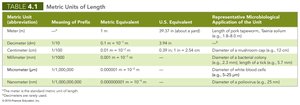
Length is measured in meters (m), with common prefixes such as kilo- (1000x), centi- (1/100x), milli- (1/1000x), micro- (1/1,000,000x), and nano- (1/1,000,000,000x).
Volume is measured in liters (L), and mass in grams (g).
Temperature is measured in degrees Celsius (°C), with the conversion formula:
Metric Unit | Meaning of Prefix | Metric Equivalent | Representative Microbiological Application |
|---|---|---|---|
Meter (m) | — | 1 m | Length of yard, tapeworm |
Millimeter (mm) | 1/1,000 | 0.001 m | Diameter of bacterial colony |
Micrometer (µm) | 1/1,000,000 | 0.000001 m | Diameter of bacterial cells |
Nanometer (nm) | 1/1,000,000,000 | 0.000000001 m | Diameter of poliovirus |
Microscopy
Microscopy is the science of using light or electrons to magnify objects, allowing scientists to observe microorganisms invisible to the naked eye. Antoni van Leeuwenhoek pioneered this field in the 17th century.
General Principles of Microscopy
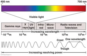
Wavelength of Radiation: The distance between two corresponding parts of a wave. Visible light ranges from 400–700 nm, and shorter wavelengths provide higher resolution.
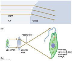
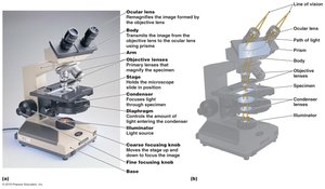
Magnification: The apparent increase in size of an object, achieved by refracting light through lenses. Total magnification is the product of the objective and ocular lens magnifications.
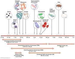
Resolution: The ability to distinguish between two points that are close together. Modern light microscopes can resolve objects as close as 0.2 µm apart.
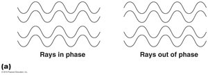
Contrast: The difference in intensity between an object and its background. Staining and phase techniques enhance contrast.
Contrast and Phase
Contrast is crucial for distinguishing microorganisms from their background. Most microbes are colorless, so staining or using light in phase increases visibility.
Types of Light Microscopy
Several types of microscopes use visible light to examine specimens, each with unique advantages for different applications.
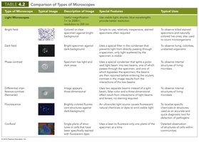
Bright-Field Microscopes
Simple: Single lens, used by Leeuwenhoek.
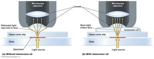
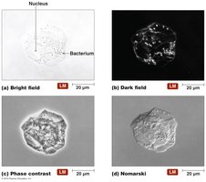
Compound: Multiple lenses, typically with 4x, 10x, 40x, and 100x (oil immersion) objectives. Oil immersion increases resolution by reducing light refraction.
Dark-Field Microscopes
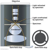
Best for observing pale or unstained specimens. A dark-field stop blocks direct light, so only scattered light enters the objective, making specimens appear bright against a dark background.
Phase Microscopes
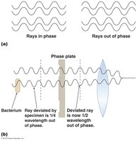
Used to view living organisms without staining. They exploit differences in refractive index to produce contrast.
Phase-Contrast: Highlights fine structures in living cells.
Differential Interference Contrast (Nomarski): Produces 3D or shadowed images using prisms.
Fluorescence Microscopes
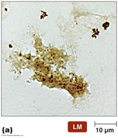
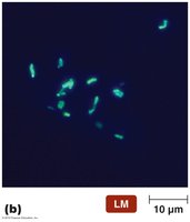
Use UV light to excite fluorescent dyes or naturally fluorescent specimens. Useful for identifying pathogens and visualizing proteins via immunofluorescence.
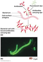
Confocal Microscopes
Use UV lasers to scan specimens stained with fluorescent dyes, producing high-resolution optical slices that can be reconstructed into 3D images. Especially useful for studying biofilms.
Electron Microscopy
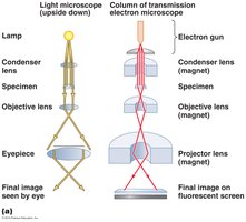
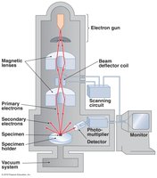
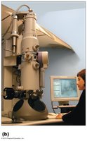
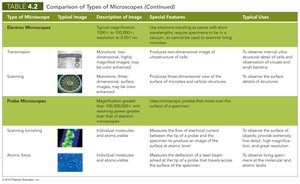
Electron microscopes use electron beams instead of light, achieving much higher magnification and resolution (down to 0.5 nm). Only dead specimens can be viewed.
Transmission Electron Microscope (TEM): Electrons pass through thin specimens, producing 2D images of internal structures.
Scanning Electron Microscope (SEM): Electrons scan the surface, producing detailed 3D images of specimen surfaces.
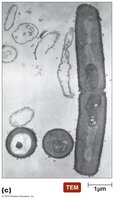
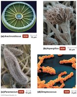
Probe Microscopy
Probe microscopes, such as Scanning Tunneling Microscopes (STM) and Atomic Force Microscopes (AFM), use physical probes to scan specimen surfaces, achieving magnifications over 100 million times and imaging at the atomic level.
STM: Measures electron tunneling between probe and specimen.
AFM: Measures deflection of a probe across the specimen surface, suitable for living cells.
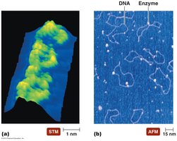
Comparison of Microscope Types
The following tables summarize the main types of microscopes, their images, features, and uses.
Staining
Principles of Staining
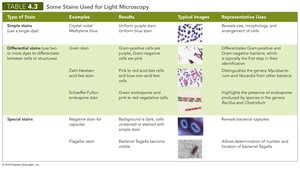
Staining enhances contrast between microorganisms and their background, making them easier to observe. Stains can be basic (cationic) or acidic (anionic), binding to different cellular components based on charge.
Simple stains: Use a single dye to reveal cell shape, size, and arrangement.
Differential stains: Use multiple dyes to distinguish between cell types or structures (e.g., Gram stain, acid-fast stain).
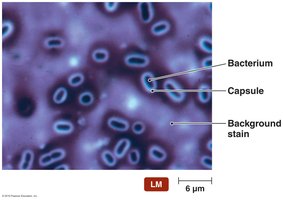
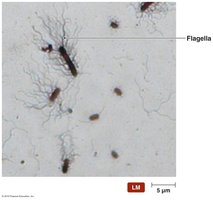
Special stains: Highlight specific structures (e.g., capsule, flagella).
Gram Stain
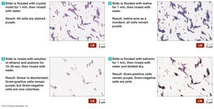
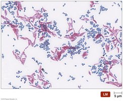
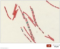
The Gram stain differentiates bacteria into Gram-positive (purple) and Gram-negative (pink) based on cell wall structure. It is a critical diagnostic tool in microbiology.
Steps: Crystal violet (primary stain), iodine (mordant), alcohol (decolorizer), safranin (counterstain).
Gram-positive: Retain crystal violet and appear purple.
Gram-negative: Lose crystal violet, take up safranin, and appear pink.
Special Stains
Negative (Capsule) Stain: Stains the background, leaving capsules clear.
Flagellar Stain: Visualizes bacterial flagella by coating them with dye and mordant.
Classification and Identification of Microorganisms
Taxonomy
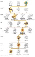
Taxonomy is the science of classifying organisms into groups (taxa) based on similarities. It includes classification, nomenclature, and identification. Modern taxonomy reflects evolutionary relationships (phylogeny) and uses genetic data.
Linnaean System: Organizes life into hierarchical categories: Domain, Kingdom, Phylum, Class, Order, Family, Genus, Species.
Binomial Nomenclature: Each organism has a two-part name: Genus (capitalized) and species (lowercase), e.g., Homo sapiens.
Three Domains: Eukarya, Bacteria, Archaea (based on rRNA sequences).
Methods of Classification and Identification
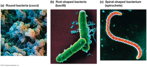
Physical Characteristics: Morphology (shape, size, arrangement) is used for identification, especially for protozoa, fungi, and bacteria.
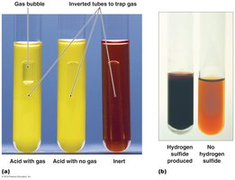
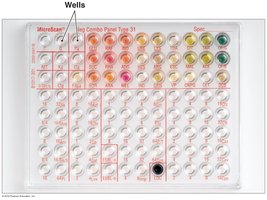
Biochemical Tests: Assess metabolic capabilities, such as carbohydrate fermentation or enzyme production.
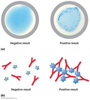
Serological Tests: Detect specific antibodies or antigens in serum, often using agglutination reactions.
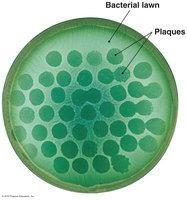
Phage Typing: Identifies bacteria based on susceptibility to specific bacteriophages.
Analysis of Nucleic Acids: Compares DNA or RNA sequences, such as G+C content, to determine relatedness.
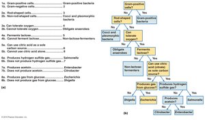
Taxonomic (Dichotomous) Keys: Stepwise tools for identifying organisms based on paired statements.
Additional info: Modern microbial taxonomy increasingly relies on molecular techniques, such as 16S rRNA sequencing, for precise identification and classification.
