 Back
BackMicroscopy, Staining, and Classification in Microbiology
Study Guide - Smart Notes
 Tailored notes based on your materials, expanded with key definitions, examples, and context.
Microscopy in Microbiology
Historical Development of Microscopy
The development of microscopy was crucial for the advancement of microbiology. Early pioneers such as Anton van Leeuwenhoek (1632-1723) created the simple microscope, allowing the first observations of microorganisms. Later, Joseph Jackson Lister (1830) developed the functional compound microscope, which improved magnification and resolution.
Structure and Function of the Modern Compound Microscope
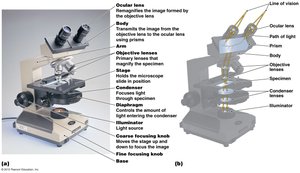
The modern compound microscope is an essential tool in microbiology, enabling the visualization of microorganisms at high magnification and resolution. It consists of several key components, each with a specific function:
Ocular lens (eyepiece): Remagnifies the image formed by the objective lens.
Objective lenses: Primary lenses that magnify the specimen.
Stage: Holds the microscope slide in position.
Condenser: Focuses light through the specimen.
Illuminator: Light source for the microscope.
Coarse and fine focusing knobs: Used to bring the specimen into sharp focus.
Relative Sizes of Microorganisms
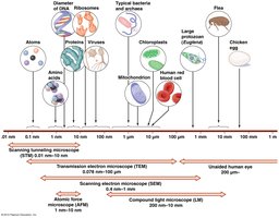
Microorganisms vary greatly in size. Typical bacteria range from 0.5–2 µm, while eukaryotic cells are larger, about 7–10 µm. Viruses, proteins, and ribosomes are much smaller and require higher magnification to be visualized.
Resolution in Microscopy
Resolution is the ability to distinguish between two points as separate entities. Higher resolution allows for the observation of finer details in specimens. The resolving power of a microscope depends on the wavelength of the illumination source and the quality of the lenses.
Light Microscopy Techniques
Bright-Field Microscopy
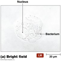
This is the most common type of light microscopy. It uses light from the base to illuminate the specimen, which appears dark against a bright background. It is suitable for stained or naturally pigmented specimens.
Oil Immersion Technique
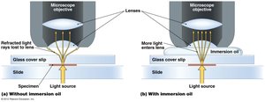
Oil immersion is used to increase the resolution of the microscope at high magnifications. A drop of immersion oil is placed between the objective lens and the slide, reducing light refraction and allowing more light to enter the lens.
Dark-Field Microscopy
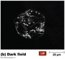
In dark-field microscopy, the specimen appears bright against a dark background. This technique is useful for observing live, unstained specimens and very pale organisms. Light is directed at an angle, and only scattered light enters the objective lens.
Phase Contrast Microscopy
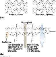
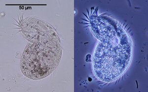
Phase contrast microscopy enhances the contrast of transparent specimens without the need for staining. It works by exploiting differences in the refractive index of cellular components, making internal structures visible in living cells.
Electron Microscopy
Principles and Types
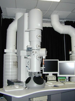
Electron microscopy uses a beam of electrons instead of light to achieve much higher resolution, allowing visualization of structures as small as 0.3 nm. There are two main types:
Transmission Electron Microscopy (TEM): Used to view internal structures of cells by transmitting electrons through thin specimens.
Scanning Electron Microscopy (SEM): Used to view surface structures, producing three-dimensional images.
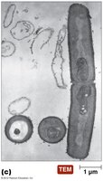
Transmission Electron Microscopy (TEM)
TEM provides detailed images of the internal structure of cells and organelles by passing electrons through ultra-thin sections of specimens.
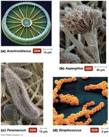
Scanning Electron Microscopy (SEM)
SEM scans the surface of a specimen with a focused beam of electrons, producing detailed three-dimensional images of surface topography.
Advantages and Disadvantages of Electron Microscopy
Advantages: Extremely high resolution and magnification; can reveal ultrastructural details.
Disadvantages: Requires vacuum conditions, complex specimen preparation, and expensive equipment.
Sample Preparation and Staining Techniques
Sample Preparation
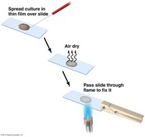
Proper sample preparation is essential for microscopy. The typical steps include:
Spreading a thin film (smear) of the specimen on a slide.
Allowing the smear to air dry.
Heat fixing the slide by passing it through a flame to adhere the cells and kill them.
Staining Techniques
Staining increases contrast and allows for the differentiation of microorganisms. There are three main types of stains:
Simple Stain: Uses one dye to color all cells, revealing general morphology but not differences between cell types.
Differential Stain: Uses multiple dyes to distinguish between different groups of organisms (e.g., Gram stain, Acid-fast stain).
Special Stains: Used to visualize specific structures such as capsules or endospores.
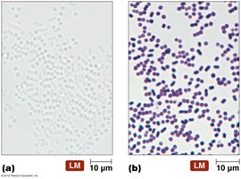
Simple Stain
A simple stain, such as crystal violet, colors all cells and allows for the observation of cell shape and arrangement.
Differential Stains: Gram Stain
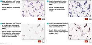
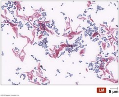
The Gram stain is the most important differential stain in microbiology. It differentiates bacteria into Gram-positive (purple) and Gram-negative (pink) based on differences in cell wall structure. The steps are:
Application of crystal violet (primary stain)
Addition of iodine (mordant)
Alcohol wash (decolorization)
Counterstain with safranin
Acid-Fast (Ziehl-Neelsen) Stain
This stain is used for bacteria with waxy cell walls (e.g., Mycobacterium and Nocardia). Acid-fast bacteria appear red, while non-acid-fast bacteria appear blue.
Special Stains
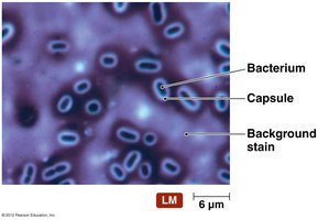
Capsule Stain (Negative Stain): Visualizes the gelatinous capsule surrounding some bacteria.
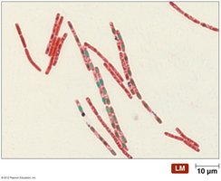
Endospore Stain: Used to detect endospores in genera such as Clostridium and Bacillus.
Bacterial Morphology and Classification
Major Bacterial Shapes
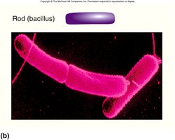
Bacteria are classified by their morphology (shape and arrangement):
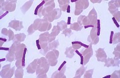
Bacillus (rod-shaped): Rigid rods, may occur singly or in chains.
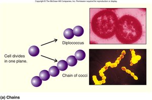
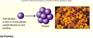
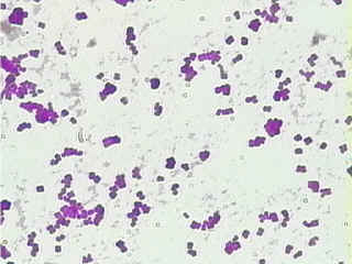
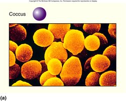
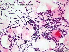
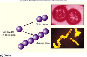
Coccus (spherical): Spherical cells, may occur singly, in pairs (diplococci), chains (streptococci), or clusters (staphylococci).
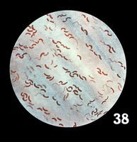
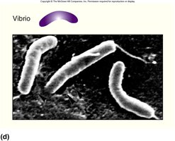
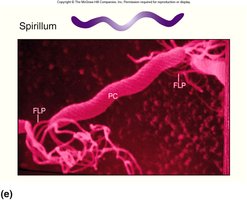
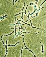
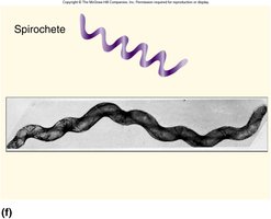
Spiral: Includes vibrio (comma-shaped), spirillum (rigid spiral), and spirochete (flexible spiral).
Spiral Bacteria
Vibrio: Comma-shaped bacteria.
Spirillum: Rigid, spiral-shaped bacteria.
Spirochete: Flexible, corkscrew-shaped bacteria.
Summary Table: Bacterial Shapes and Arrangements
Shape | Arrangement | Example |
|---|---|---|
Bacillus (rod) | Single, chains (streptobacillus) | Bacillus subtilis |
Coccus (spherical) | Single, pairs (diplococcus), chains (streptococcus), clusters (staphylococcus), packets | Staphylococcus aureus, Streptococcus pyogenes |
Vibrio | Single | Vibrio cholerae |
Spirillum | Single | Spirillum volutans |
Spirochete | Single | Treponema pallidum |
Additional info: Understanding bacterial morphology and staining techniques is essential for identification and classification in clinical and research microbiology. Mastery of microscopy and staining is foundational for all microbiologists.
