Back
BackMicroscopy, Staining, and Classification in Microbiology
Study Guide - Smart Notes
 Tailored notes based on your materials, expanded with key definitions, examples, and context.
Microscopy: How We See the Invisible World
Introduction to Microscopy
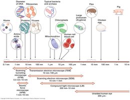
Microscopy is the technology that allows us to visualize objects too small to be seen by the naked eye, such as microorganisms. By using light or electrons, microscopes magnify objects and alter the direction of light rays, presenting them at a greater angle of vision. This makes small objects appear much larger and more detailed to the observer.
General Principles of Microscopy
Magnification: The apparent increase in size of an object, indicated by a number and “X” (e.g., 100X means 100 times larger).
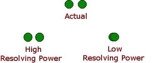
Resolution: The ability to distinguish between two objects that are close together. High resolution allows for clearer, more detailed images.
Resolving Power: Inversely proportional to the wavelength of the radiation used (light or electrons). Shorter wavelengths provide higher resolving power.
Contrast in Microscopy
Contrast is the difference between the object and the background. It can be improved by using dyes or manipulating light, making it easier to distinguish cells from their surroundings.
Types of Microscopes
Light Microscopes
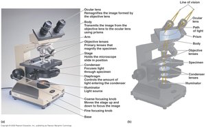
Bright-Field Microscopes: Use visible light to illuminate specimens. Simple versions have a single lens (as used by Leeuwenhoek), while compound microscopes use multiple lenses for greater magnification and resolution.
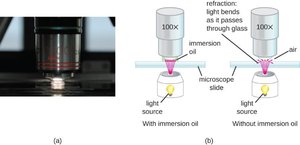
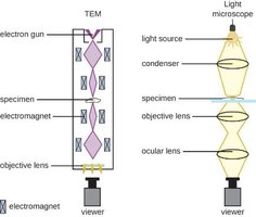
Compound Microscopes: Feature a series of lenses, including objective and ocular lenses. Oil immersion lenses increase resolution by reducing light refraction. Total magnification is the product of the objective and ocular lens magnifications.


Electron Microscopes
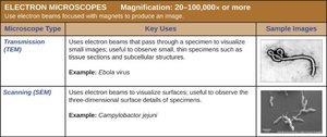
Electron microscopes use beams of electrons instead of light, allowing for much higher magnification and resolution. They can visualize structures as small as molecules and large atoms.
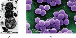
Transmission Electron Microscope (TEM): Used to study internal cell structures by passing electrons through thin specimen sections. Produces 2-D images.
Scanning Electron Microscope (SEM): Used to study surface architecture by scanning the specimen with electrons. Produces 3-D images.
Techniques for Identifying Microorganisms
Physical Characteristics
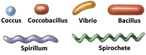
Morphology: Shape and arrangement of cells (e.g., coccus, bacillus, spirillum).
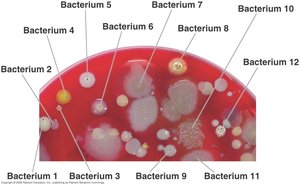
Colony Appearance: The look of bacterial colonies on agar plates.
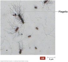
Identifying Features: Presence of endospores, flagella, or capsules.
Biochemical Tests
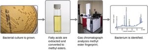
Biochemical tests assess the ability of microorganisms to use or produce specific chemicals, such as fermenting carbohydrates or producing hydrogen sulfide gas. Fatty acid composition can also be analyzed using FAME (Fatty Acid Methyl Ester) analysis, which produces a unique chromatogram for each bacterium.
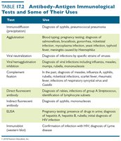
Serological Tests
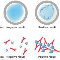
Antibodies: Immune proteins that bind to specific antigens on microorganisms, causing them to clump (agglutinate).
Serology: The study of antigen-antibody interactions in the lab.
Antiserum: Solution containing antibodies used to identify pathogens.
Agglutination Test: Mixing antisera with a sample to detect clumping, indicating the presence of the target antigen.
Staining Techniques
Principles of Staining
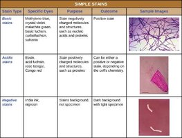
Staining increases contrast, making cells visible against the background. Most stains are either cationic (basic, positively charged) or anionic (acidic, negatively charged). Basic stains bind to negatively charged cell components, while acidic stains bind to positively charged components.
Simple Stain
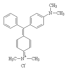
Uses a single dye (e.g., methylene blue, crystal violet).
Reveals basic cell size, shape, and arrangement.
All cells stain similarly due to overall charge interactions.
Differential Stains
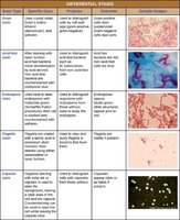
Differential stains use two or more dyes to distinguish between different types of cells or cell structures. Common examples include the Gram stain, acid-fast stain, and endospore stain.
Gram Stain
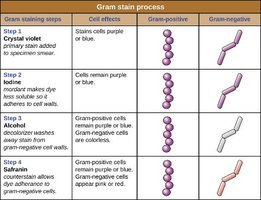
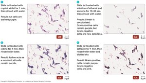
Gram-positive cells: Thick peptidoglycan layer, retain crystal violet, appear purple.
Gram-negative cells: Thin peptidoglycan layer, lose crystal violet after decolorization, appear pink after counterstaining with safranin.
Acid-Fast Stain
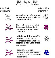
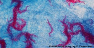
Used for bacteria with waxy mycolic acid in their cell walls (e.g., Mycobacterium).
Primary stain: carbolfuchsin (red), heat as mordant.
Decolorizer: acid-alcohol; non-acid-fast cells become colorless.
Counterstain: methylene blue; non-acid-fast cells turn blue, acid-fast remain red.
Endospore Stain
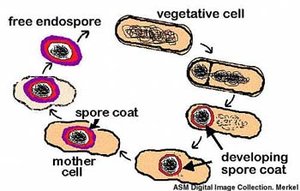
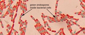
Differentiates endospore-forming bacteria (e.g., Bacillus, Clostridium).
Primary stain: malachite green with heat.
Decolorizer: water; only endospores retain green.
Counterstain: safranin; vegetative cells turn pink, endospores remain green.
Negative Stain
Uses acidic dyes (e.g., nigrosine, India ink) that stain the background, leaving cells colorless.
Useful for visualizing capsules and cell morphology.
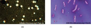
Capsule Stain
Combines negative staining (background) with simple staining (cell).
Capsules appear as clear halos around cells.
Flagella Stain
Special stains bind to and thicken flagella, making them visible under the light microscope.
Used to determine motility and arrangement of flagella.
Specimen Preparation for Light Microscopy
Wet Mounts
A drop of medium containing organisms is placed on a slide for immediate observation.
Carboxymethylcellulose can be added to slow movement of fast organisms.
Smears and Heat Fixation
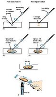
Microorganisms are spread on a slide, air-dried, and heat-fixed by passing through a flame or using a slide warmer.
Heat fixation kills organisms, adheres them to the slide, and makes them more receptive to stains.