Back
Microscopy, Staining, and Classification in Microbiology
Study Guide - Smart Notes
 Tailored notes based on your materials, expanded with key definitions, examples, and context.
Microscopy in Microbiology
Electron Microscopy: Principles and Applications
Electron microscopy is a powerful technique that uses electron beams instead of light to visualize specimens, allowing for much higher resolution and magnification than light microscopy. This enables detailed observation of bacteria, viruses, cellular structures, and even large molecules.
Resolution: Electron beams have wavelengths between 0.01 nm and 0.001 nm, compared to visible light's 400 nm, allowing electron microscopes to resolve much smaller structures.
Magnification: Electron microscopes can magnify objects from 10,000x to over 100,000x.
Applications: Used to study bacteria, viruses, internal cell structures, and macromolecules.
Types of Electron Microscopes
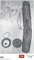
Transmission Electron Microscope (TEM): Electrons pass through thin specimens, revealing internal structures in great detail.
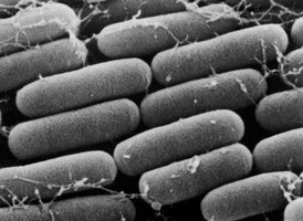
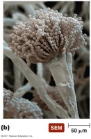
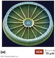
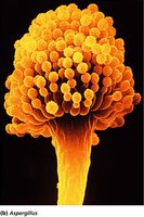
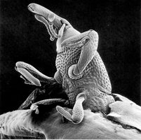
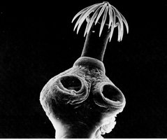
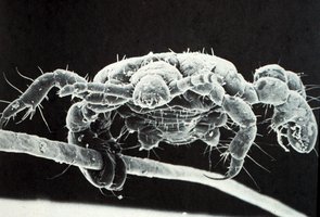
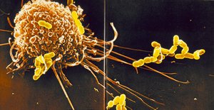
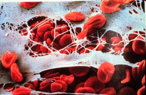
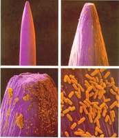
Scanning Electron Microscope (SEM): Electrons scan the surface of specimens, producing detailed 3D images of surfaces.
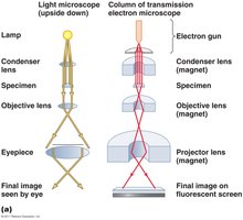
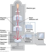
Examples of Electron Microscopy Images
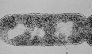
TEM Images: Show internal details of cells and viruses.
SEM Images: Reveal surface structures of microorganisms and small organisms.

Staining Techniques in Microbiology
Purpose and Principles of Staining
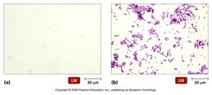
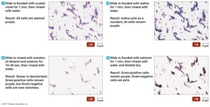
Staining increases the contrast and resolution of microscopic specimens, making cellular structures more visible. Microbiological stains are typically salts with a colored ion (chromophore). The choice of stain depends on the chemical properties of the specimen.
Heat-fixing: Fixes and kills organisms on the slide, preserving their structure for staining.
Acidic dyes: Stain alkaline (basic) structures.
Basic dyes: Stain acidic (negatively charged) structures; most commonly used for bacteria.
Types of Staining Procedures
Simple Stains: Use a single dye to highlight the entire organism.
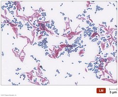
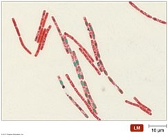
Differential Stains: Distinguish between different types of organisms or structures (e.g., Gram stain, acid-fast stain, endospore stain).
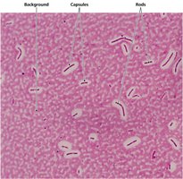
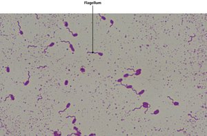
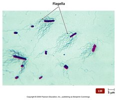
Special Stains: Highlight specific structures (e.g., capsule, flagella, fluorescent stains).
Staining for Electron Microscopy: Uses heavy metal salts to increase electron density and contrast.
Classification and Identification of Microorganisms
Taxonomy: Principles and Importance
Taxonomy is the science of classification, nomenclature, and identification of organisms. It organizes biological diversity and reflects evolutionary relationships, enabling scientists to predict characteristics and behaviors of organisms based on relatedness.
Classification: Grouping organisms based on shared characteristics.
Nomenclature: Assigning standardized names (binomial nomenclature).
Identification: Determining the identity of an organism.
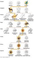
Carl Linnaeus established the modern system of taxonomy, grouping similar organisms into species and higher categories. Modern taxonomy emphasizes phylogenetic relationships, often using genetic data.
Modern Taxonomic Systems
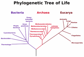
Domains: Based on ribosomal RNA sequences, Carl Woese proposed three domains: Bacteria, Archaea, and Eukarya.
Phylogenetic Hierarchy: Reflects evolutionary relationships among organisms.
Methods for Identifying Microorganisms
Physical Characteristics: Morphology, staining properties, and colony appearance.
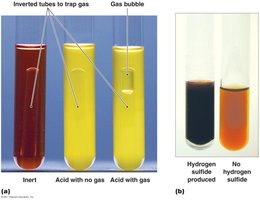
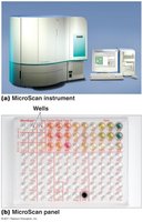
Biochemical Tests: Metabolic capabilities, such as fermentation and enzyme production.
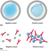
Serological Tests: Use antibodies to detect specific microbial antigens.
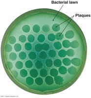
Phage Typing: Uses bacteriophages to identify bacterial strains.
MALDI-TOF Mass Spectrometry: Identifies organisms based on protein profiles.
Nucleic Acid Analysis: PCR and sequencing to detect and classify organisms based on genetic material.
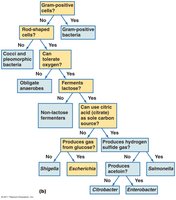
Taxonomic Keys
Dichotomous keys are tools that guide users through a series of paired statements to identify organisms. Each choice leads to another pair of statements or to the identification of the organism.