 Back
BackChapter Four: Microscopy, Staining, and Classification
Study Guide - Smart Notes
 Tailored notes based on your materials, expanded with key definitions, examples, and context.
Microscopy, Staining, and Classification
Introduction
This chapter explores the fundamental principles and applications of microscopy, staining, and the classification of microorganisms. Mastery of these topics is essential for understanding how microbiologists observe, identify, and categorize microbes in clinical and research settings.
Units of Measurement in Microbiology
Metric Units and Their Relevance
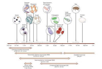
Microbiologists use the metric system to measure microorganisms and their components due to its universal standardization and ease of conversion. The most common units include meters, millimeters, micrometers, and nanometers.
Meter (m): The base unit of length in the metric system.
Micrometer (µm): 1 µm = 10-6 m; used to measure bacteria.
Nanometer (nm): 1 nm = 10-9 m; used to measure viruses and molecular structures.
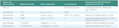
Metric Unit | Meaning of Prefix | Metric Equivalent | U.S. Equivalent | Representative Microbiological Application |
|---|---|---|---|---|
Meter (m) | — | 1 m | 39.37 in. (about 1 yard) | Length of tape measures, S. aureus colony (1–1.5 mm) |
Decimeter (dm) | 1/10 | 0.1 m = 10-1 m | 3.94 in. | Diameter of a watermelon |
Centimeter (cm) | 1/100 | 0.01 m = 10-2 m | 0.39 in. = 2.54 cm | Diameter of a chicken egg (about 3–5 cm) |
Millimeter (mm) | 1/1,000 | 0.001 m = 10-3 m | 0.039 in. | Diameter of a red blood cell (about 7–8 µm) |
Micrometer (µm) | 1/1,000,000 | 0.000001 m = 10-6 m | 0.000039 in. | Diameter of white blood cells (about 8–25 µm) |
Nanometer (nm) | 1/1,000,000,000 | 0.000000001 m = 10-9 m | 0.000000039 in. | Diameter of a poliovirus (about 25 nm) |
Principles of Microscopy
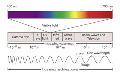
Electromagnetic Spectrum and Wavelength
Microscopes utilize different types of electromagnetic radiation, including visible light and ultraviolet (UV) light. The wavelength of the radiation used affects the resolving power of the microscope; shorter wavelengths provide better resolution.
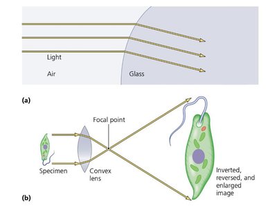
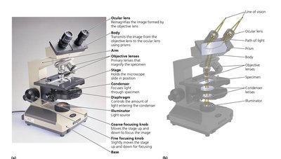
Magnification and Image Formation
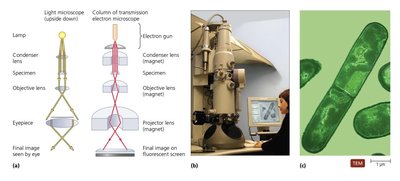
Magnification is the process of enlarging the appearance of an object. Convex glass lenses bend (refract) light to focus and magnify images, producing an inverted and reversed image of the specimen.
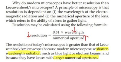
Resolution
Resolution is the ability to distinguish two points as separate entities. The limit of resolution (d) is the minimum distance at which two points can be distinguished. A lower value of d indicates better resolution. The formula for resolution is:
Modern microscopes achieve better resolution by using shorter-wavelength radiation and lenses with larger numerical apertures.
Comparative Resolving Power
Different microscopes and the human eye have varying resolving powers, which determine the smallest objects they can distinguish. Electron microscopes have much greater resolving power than light microscopes.
Contrast
Contrast is the difference in intensity between an object and its background. Increasing contrast, often by staining, improves resolution and clarity.
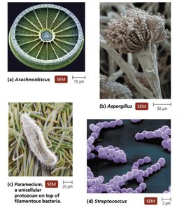
Types of Microscopes
Light Microscopy
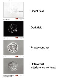
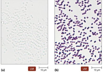
Bright-Field Microscopes: Use visible light to illuminate specimens; stained specimens appear against a bright background. Compound microscopes use multiple lenses for higher magnification.
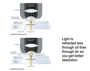
Oil Immersion: Using oil between the slide and lens increases resolution by reducing light refraction.
Dark-Field Microscopes: Best for observing pale or colorless objects; only scattered light enters the objective lens, making specimens appear bright against a dark background.
Phase Microscopes: Enhance contrast in transparent specimens without staining. Includes phase-contrast and differential interference contrast (DIC) microscopes.
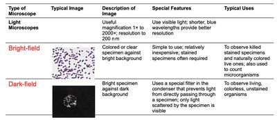
Comparison of Light Microscopy Types
Type | Description | Special Features | Typical Uses |
|---|---|---|---|
Bright-field | Colored or clear specimen against bright background | Simple to use; stained specimens often required | To observe stained specimens and naturally colored organisms |
Dark-field | Bright specimen against dark background | Special filter blocks direct light | To observe living, colorless, unstained organisms |
Phase-contrast | Specimen has light and dark areas | Special condenser splits light beam | To observe internal structures of living microbes |
DIC (Nomarski) | Image appears three-dimensional | Uses two beams; no staining required | To observe internal structures of living microbes |
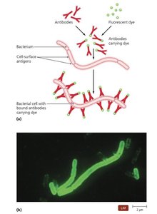
Fluorescence and Confocal Microscopy
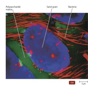
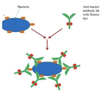
Fluorescence Microscopes: Use UV light to excite fluorescent dyes or naturally fluorescent specimens, increasing resolution and contrast. Used in immunofluorescence to detect specific pathogens.
Confocal Microscopes: Use lasers to illuminate a single plane of a specimen stained with fluorescent dyes, producing high-resolution, three-dimensional images.
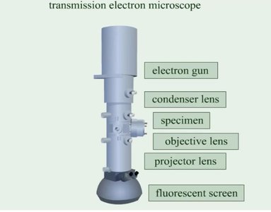
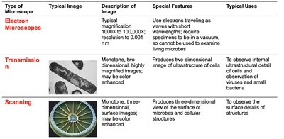
Electron Microscopy
Electron microscopes use electron beams instead of light, providing much higher magnification and resolution. Two main types are:
Transmission Electron Microscope (TEM): Produces two-dimensional images of internal structures.
Scanning Electron Microscope (SEM): Produces three-dimensional images of specimen surfaces.
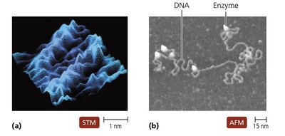
Probe Microscopy
Probe microscopes, such as scanning tunneling and atomic force microscopes, can visualize individual molecules and atoms by scanning the surface with a physical probe.
Staining Techniques
Principles and Preparation
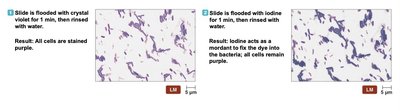
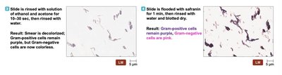
Staining increases contrast and resolution, making microorganisms easier to observe under a microscope. Dyes are usually salts with a colored ion (chromophore). Basic dyes stain acidic structures, while acidic dyes stain basic structures.
Types of Stains
Simple Stains: Use a single dye to reveal cell shape, size, and arrangement.
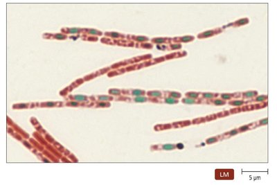
Differential Stains: Use multiple dyes to distinguish between different types of cells or structures. Common examples include Gram stain, acid-fast stain, and endospore stain.
Special Stains: Used to highlight specific structures such as capsules (negative stains) or flagella.
Classification and Identification of Microorganisms
Taxonomy and Nomenclature
Taxonomy is the science of classifying organisms, involving classification, nomenclature, and identification. The modern system reflects evolutionary relationships and includes three domains: Eukarya, Bacteria, and Archaea.
Binomial Nomenclature: Each organism is given a two-part scientific name (genus and species).
Taxonomic Hierarchy: Domain, Kingdom, Phylum, Class, Order, Family, Genus, Species.
Methods of Identification
Physical Characteristics: Morphology of cells and colonies.
Biochemical Tests: Assess metabolic capabilities, such as sugar fermentation or enzyme production.
Serological Tests: Detect antigen-antibody reactions to identify specific microbes.
Phage Typing: Uses bacteriophages to determine bacterial identity based on susceptibility to infection.
Analysis of Nucleic Acids: DNA or RNA sequencing for precise classification.
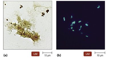
Clinical Application Example: Lyme Disease Diagnosis
Immunofluorescence microscopy can be used to detect Borrelia burgdorferi in blood samples by tagging specific antibodies with fluorescent dyes. Serological tests, such as agglutination assays, use antibodies to detect the presence of bacterial antigens in patient serum.
Summary Table: Types of Microscopes
Type | Description | Special Features | Typical Uses |
|---|---|---|---|
Light Microscopes | 1x to 2000x; resolution to 200 nm | Visible light; blue wavelengths better | Stained specimens, cell counting |
Electron Microscopes | 1000x to 100,000x; resolution to 0.001 nm | Electrons; vacuum required | Ultrastructure of cells, viruses |
Probe Microscopes | 100,000,000x+; atomic resolution | Physical probes | Molecules, atoms |
Key Takeaway: Mastery of microscopy, staining, and classification techniques is foundational for the observation, identification, and study of microorganisms in microbiology.
