 Back
BackMicroscopy, Staining, and Classification in Microbiology CH4-1
Study Guide - Smart Notes
 Tailored notes based on your materials, expanded with key definitions, examples, and context.
Microscopy, Staining, and Classification
Overview of Microscopy and Staining
Microscopy and staining are foundational techniques in microbiology, enabling scientists to observe, differentiate, and classify microorganisms. Understanding the principles of microscopy, the use of stains, and the classification systems is essential for identifying and studying microbes.
Units of Measurement in Microbiology
Metric Units and Their Application
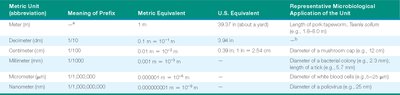
Metric System: Scientists use the metric system for consistency and ease of conversion. The meter (m) is the base unit, with subunits such as millimeter (mm), micrometer (µm), and nanometer (nm) commonly used in microbiology.
Application: Microbiologists use these units to measure cells, viruses, and cellular structures.
Metric Unit | Meaning of Prefix | Metric Equivalent | Representative Microbiological Application |
|---|---|---|---|
Meter (m) | — | 1 m | Length of tapeworm |
Millimeter (mm) | 1/1,000 | 0.001 m | Diameter of bacterial colony |
Micrometer (µm) | 1/1,000,000 | 0.000001 m | Diameter of white blood cell |
Nanometer (nm) | 1/1,000,000,000 | 0.000000001 m | Diameter of poliovirus |
Principles of Microscopy
Wavelength, Magnification, Resolution, and Contrast
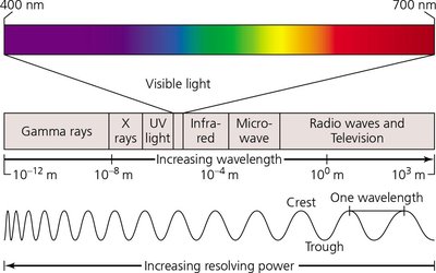
Wavelength: The distance between two corresponding parts of a wave. Shorter wavelengths provide higher resolution.
Magnification: The apparent increase in size of an object. Calculated as the product of the magnifications of the objective and ocular lenses.
Resolution: The ability to distinguish two points as separate entities. Higher resolution allows for clearer images of small structures.
Contrast: The difference in intensity between an object and its background. Staining and phase techniques enhance contrast.
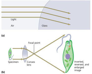
Light Refraction and Image Formation
Light bends (refracts) as it passes through different media, such as air and glass.
Convex lenses focus light to magnify specimens, producing inverted and enlarged images.
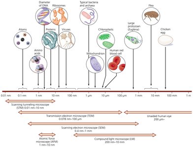
Limits of Resolution
The human eye, light microscopes, and electron microscopes have different resolving powers, determining the smallest objects they can distinguish.
Electron microscopes can resolve much smaller structures than light microscopes.
Types of Microscopy
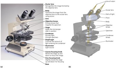
Light Microscopy
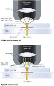
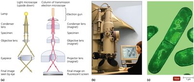
Bright-Field Microscopes: Use visible light to illuminate specimens. Simple (single lens) and compound (multiple lenses) types exist. Oil immersion increases resolution by reducing light refraction.
Dark-Field Microscopes: Enhance contrast for pale or colorless specimens by only collecting light scattered by the specimen, making it appear bright against a dark background.
Phase Microscopes: Increase contrast by exploiting differences in refractive index. Includes phase-contrast and differential interference contrast (Nomarski) microscopes, useful for observing living cells.

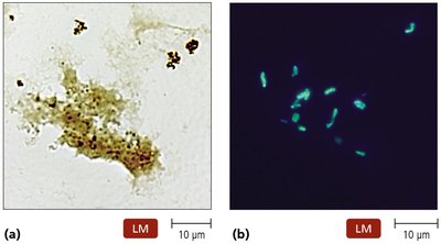
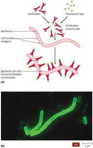
Fluorescence Microscopes: Use UV light to excite fluorescent dyes or naturally fluorescent specimens, increasing resolution and contrast. Widely used in immunofluorescence for pathogen detection.
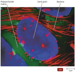
Confocal Microscopes: Use lasers and fluorescent dyes to produce sharp, three-dimensional images by focusing on a single plane within the specimen.
Electron Microscopy
Electron microscopes use electron beams instead of light, achieving much higher magnification and resolution.
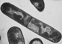
Transmission Electron Microscopes (TEM): Produce detailed two-dimensional images of internal cell structures.
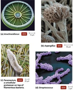
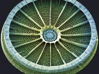
Scanning Electron Microscopes (SEM): Generate three-dimensional images of specimen surfaces.
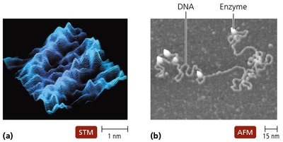
Probe Microscopy
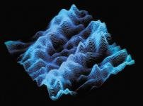
Probe microscopes, such as scanning tunneling microscopes (STM) and atomic force microscopes (AFM), can visualize surfaces at the atomic level.
Comparison of Microscopes
Type | Image | Special Features | Typical Uses |
|---|---|---|---|
Bright-field | Colored/clear specimen on bright background | Simple, inexpensive, often requires staining | Observe killed, stained specimens |
Dark-field | Bright specimen on dark background | Special condenser, no direct light | Observe living, colorless organisms |
Phase-contrast | Light and dark areas | Special condenser splits light beam | Observe internal structures of living microbes |
Fluorescence | Bright fluorescent structures on dark background | UV light source, fluorescent dyes | Detect pathogens, localize chemicals |
Confocal | Single plane, fluorescently stained | Laser illumination, 3D imaging | Detailed cell structure observation |
TEM | 2D, highly magnified | Electrons, vacuum required | Internal cell details, viruses |
SEM | 3D, surface images | Electrons, vacuum required | Surface details |
STM/AFM | Atomic/molecular detail | Microscopic probes | Surface, molecular, atomic level |
Staining Techniques
Principles of Staining
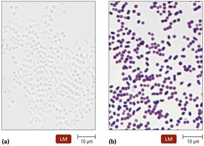
Staining increases contrast and resolution, making microorganisms easier to observe under a microscope.
Dyes are usually salts with a colored ion (chromophore).
Acidic dyes stain alkaline structures; basic dyes stain acidic structures (most cells are negatively charged, so basic dyes are more common).
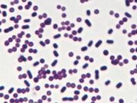
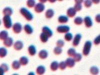
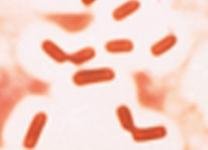
Simple Stains
Use a single basic dye (e.g., crystal violet, safranin, methylene blue).
Reveal cell size, shape, and arrangement.
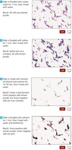
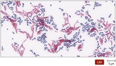
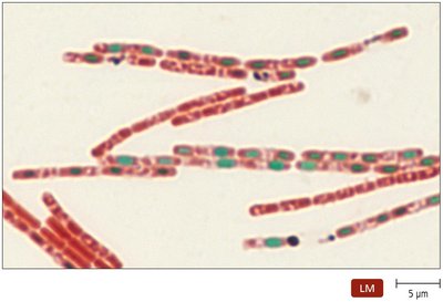
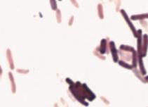
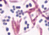
Differential Stains
Use two or more dyes to distinguish between cell types or structures.
Common types: Gram stain, acid-fast stain, endospore stain, histological stains.
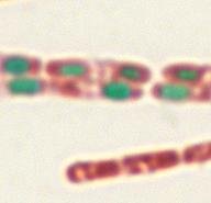
Special Stains
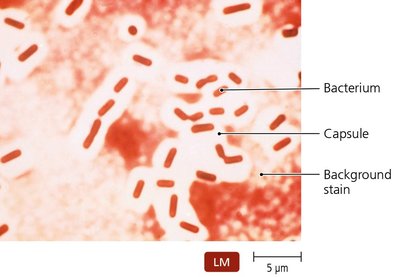
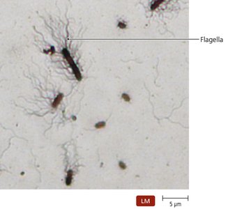
Highlight specific microbial structures, such as capsules, flagella, or use fluorescent dyes.
Negative stains color the background, leaving cells unstained.
Flagellar stains make bacterial flagella visible.
Summary Table: Stains Used in Light Microscopy
Type of Stain | Examples | Results | Representative Uses |
|---|---|---|---|
Simple | Crystal violet, methylene blue | Uniform color | Size, morphology, arrangement |
Gram | Gram stain | Purple (Gram+), pink (Gram-) | Differentiates Gram+/- bacteria |
Acid-fast | Ziehl-Neelsen | Pink/red acid-fast, blue non-acid-fast | Identifies Mycobacterium, Nocardia |
Endospore | Schaeffer-Fulton | Green endospores, pink/red cells | Detects Bacillus, Clostridium endospores |
Negative | Capsule stain | Dark background, unstained cells | Reveals capsules |
Flagellar | Flagella stain | Flagella visible | Number/location of flagella |
Staining for Electron Microscopy
Uses chemicals containing heavy metals to increase electron density and contrast.
Stains may bind to the specimen or the background.
Classification and Identification of Microorganisms
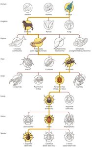
Taxonomy: Classification, Nomenclature, and Identification
Taxonomy organizes organisms into groups based on similarities, predicts characteristics, and reflects evolutionary relationships.
Linnaeus developed the binomial nomenclature system and initially proposed two kingdoms; modern taxonomy uses three domains (Eukarya, Bacteria, Archaea) based on rRNA sequences.
Taxonomic and Identifying Characteristics
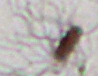
Physical Characteristics: Morphology of cells and colonies can aid identification.
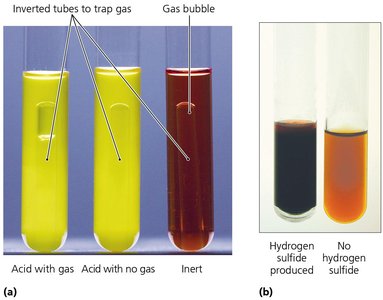
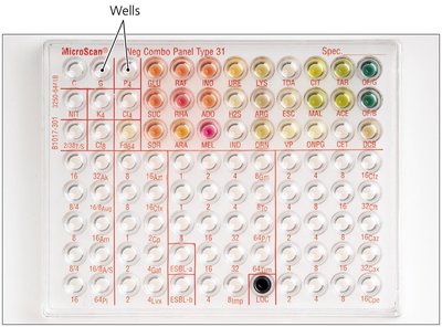
Biochemical Tests: Assess metabolic capabilities, such as sugar fermentation or enzyme production.
Serological Tests: Use antigen-antibody reactions to identify organisms.
Phage Typing: Uses bacteriophage specificity to distinguish bacterial strains.
MALDI/TOF Mass Spectrometry: Identifies microbes by their protein profiles.
Analysis of Nucleic Acids: DNA/RNA sequencing and G+C content analysis are used for classification.
Taxonomic Keys
Dichotomous keys use a series of paired statements to guide users to the identification of an organism.
Summary
Microscopy and staining are essential for visualizing and differentiating microorganisms.
Various types of microscopes and stains are used depending on the application and the structures of interest.
Classification systems help organize microbial diversity and facilitate identification in clinical and research settings.
