 Back
Microscopy, Staining, and Classification: Principles and Applications in Microbiology
Study Guide - Smart Notes
 Tailored notes based on your materials, expanded with key definitions, examples, and context.
Microscopy, Staining, and Classification
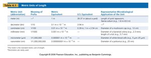
Metric Units of Length in Microbiology
Understanding metric units is essential for accurately describing the size of microorganisms and cellular structures. Microbiologists use units ranging from meters to picometers to measure and compare biological specimens.
Meter (m): The base unit of length in the metric system.
Micrometer (μm): Commonly used for bacteria and cells; 1 μm = m.
Nanometer (nm): Used for viruses and molecular structures; 1 nm = m.
Picometer (pm): Used for atomic and subatomic measurements; 1 pm = m.
Metric Unit | Meaning of Prefix | Metric Equivalent | Representative Microbiological Application |
|---|---|---|---|
meter (m) | 1 | 1 m | Length of path specimen travels |
micrometer (μm) | 1/1,000,000 | 0.000001 m | Diameter of bacteria, mitochondria |
nanometer (nm) | 1/1,000,000,000 | 0.000000001 m | Diameter of viruses, ribosomes |
picometer (pm) | 1/1,000,000,000,000 | 0.000000000001 m | Wavelength of electron beam |
Sizes of Microbiological Objects
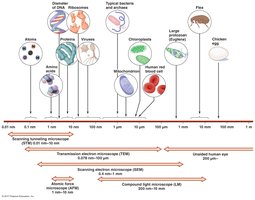
Microbiological objects vary greatly in size, from atoms and molecules to cells and multicellular organisms. Understanding these sizes is crucial for selecting appropriate microscopy techniques.
Viruses: Typically measured in nanometers.
Bacteria: Usually measured in micrometers.
Eukaryotic cells: Range from a few micrometers to hundreds of micrometers.
Principles of Microscopy
General Principles of Microscopy
Microscopy relies on four general principles: magnification, resolution, contrast, and illumination. These principles are optimized to visualize microorganisms and cellular structures.
Magnification: The process of enlarging the appearance of an object.
Resolution: The ability to distinguish two points as separate entities. Higher resolution allows for clearer images.
Contrast: The difference in light intensity between the specimen and its background, often enhanced by staining.
Illumination: The light source used to visualize the specimen.
Electromagnetic Spectrum and Resolving Power
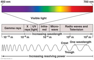
The resolving power of a microscope depends on the wavelength of light or electrons used. Shorter wavelengths provide higher resolution.
Visible light: Used in light microscopy; wavelength ranges from 400–700 nm.
UV light: Used in fluorescent microscopy; shorter wavelength increases resolution.
Electron beams: Used in electron microscopy; wavelength in picometers allows visualization of atomic structures.
Light Refraction and Magnification
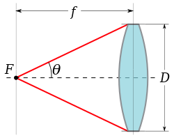
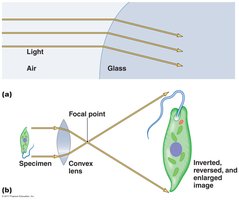
Refraction is the bending of light as it passes through different media, which is fundamental to magnification in microscopy. Lenses focus light to produce enlarged images of specimens.
Refractive index: A measure of how much a substance bends light; affects image clarity.
Convex lenses: Used to focus light and magnify specimens.
Types of Light Microscopy
Bright-Field Microscopy
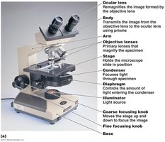
Bright-field microscopes are the most common type, used primarily for stained specimens. They can be simple (one lens) or compound (multiple lenses).
Simple microscope: Contains a single magnifying lens; used by Leeuwenhoek.
Compound microscope: Uses a series of lenses for higher magnification and resolution.
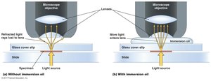
Oil immersion: Increases resolution by reducing light refraction at high magnification.
Total magnification:
Dark-Field Microscopy
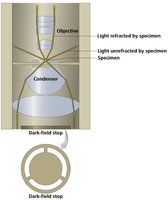
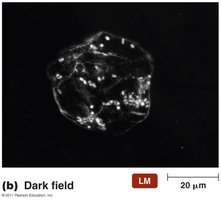
Dark-field microscopes are best for observing pale or thin living specimens. They use a dark-field stop to block direct light, allowing only scattered light to reach the objective lens.
Contrast: Specimen appears light against a dark background.
Applications: Enables observation of thin and translucent organisms.
Phase Microscopy
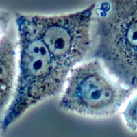
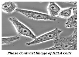
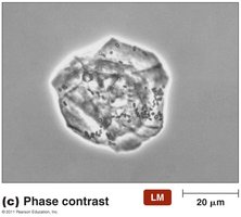
Phase microscopes are used to examine living organisms or specimens that would be damaged by staining. They create contrast by treating light rays differently based on their phase.
Phase-contrast microscope: Enhances contrast by exploiting differences in refractive index.
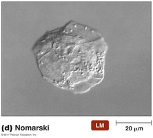
Differential Interference Contrast (DIC) microscope: Provides high-contrast, 3D-like images.
Fluorescent and Confocal Microscopy
Fluorescent Microscopy
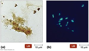
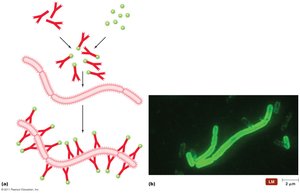
Fluorescent microscopes use UV light to excite fluorochromes, which emit visible light. This technique is valuable for identifying pathogens and visualizing cellular proteins.
Immunofluorescence: Uses antibodies labeled with fluorescent dyes to detect specific antigens.
Applications: Used in clinical diagnostics and research.
Confocal Microscopy
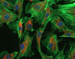
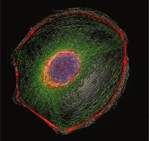
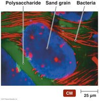
Confocal microscopes use lasers to illuminate a single plane of a specimen, increasing resolution and allowing for 3D reconstruction. Emitted light passes through a pinhole aperture, reducing background noise.
Resolution: Increased by up to 40% compared to conventional light microscopy.
3D imaging: Computer software constructs three-dimensional images from digitized data.
Summary Table: Types of Light Microscopy
Microscope Type | Principle | Best Use | Image Appearance |
|---|---|---|---|
Bright-field | Direct light through specimen | Stained specimens | Specimen dark on light background |
Dark-field | Oblique light rays, scattered by specimen | Thin, pale, living specimens | Specimen light on dark background |
Phase-contrast | Light rays out of phase | Living, unstained specimens | Enhanced contrast, internal structures visible |
DIC (Nomarski) | Interference of polarized light | 3D-like images of living cells | High-contrast, pseudo-3D |
Fluorescent | UV light excites fluorochromes | Pathogen identification, protein localization | Bright colored structures on dark background |
Confocal | Laser illumination, pinhole aperture | 3D imaging of cells and tissues | Sharp, high-resolution, 3D images |
