 Back
BackMicroscopy, Staining, and Classification: Principles and Applications in Microbiology
Study Guide - Smart Notes
 Tailored notes based on your materials, expanded with key definitions, examples, and context.
Microscopy, Staining, & Classification
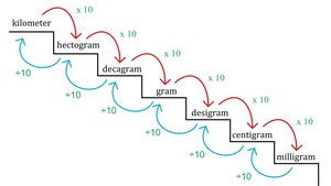
Units of Measurement in Microscopy
Understanding the scale of microorganisms is essential in microbiology. The metric system is used to measure microscopic entities, with units such as micrometers (µm) and nanometers (nm) being most common.
Micrometer (µm): 1 µm = 10-6 meters
Nanometer (nm): 1 nm = 10-9 meters
Prefixes: kilo-, centi-, milli-, micro-, nano-, pico-

General Principles of Microscopy
Microscopy is the technique used to visualize microorganisms. The main principles include wavelength, magnification, resolution, and contrast.
Wavelength: Shorter wavelengths provide higher resolution.
Magnification: The process of enlarging the appearance of an object.
Resolution: The ability to distinguish two close points as separate entities.
Contrast: The difference in brightness between the specimen and its background.
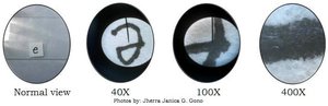
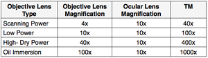
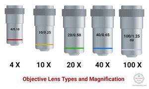
Magnification
Magnification is achieved by using lenses that bend light rays to enlarge the image of the specimen. The total magnification is the product of the objective lens and ocular lens magnifications.
Formula:
Example: 40x objective × 10x ocular = 400x total magnification
Resolution
Resolution determines the clarity and detail of the image. It depends on the wavelength of light and the numerical aperture (NA) of the lens.
Numerical Aperture (NA): A measure of a lens's ability to gather light and resolve fine specimen detail at a fixed object distance.
Formula:
Contrast and Staining
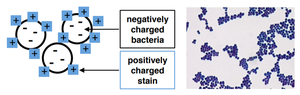
Contrast is essential for distinguishing features of microorganisms. Staining increases contrast by coloring cells or their background, making transparent specimens visible.
Staining: The process of adding dyes to specimens to enhance visibility.
Types of stains: Basic (cationic) stains color cells; acidic (anionic) stains color the background.

Types of Light Microscopy
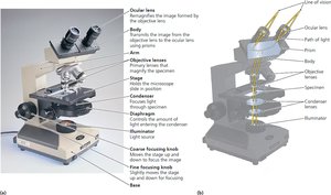
Simple and Compound Light Microscopes
Light microscopes use visible light to illuminate specimens. Simple microscopes have one lens, while compound microscopes use multiple lenses for higher magnification and resolution.
Simple microscope: Single lens system.
Compound microscope: Multiple lenses; includes objective and ocular lenses.
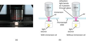
Oil immersion: Increases resolution by reducing light refraction.
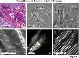
Bright-Field, Dark-Field, Phase, and Fluorescence Microscopy
Different microscopy techniques are used to enhance contrast and visualize specific features of microorganisms.
Bright-field: Standard illumination; specimen appears dark against a bright background.
Dark-field: Only scattered light enters the lens; specimen appears bright against a dark background.
Phase-contrast: Enhances contrast in transparent specimens without staining.
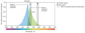
Fluorescence: Uses fluorophores that emit light upon excitation; useful for detecting specific molecules.
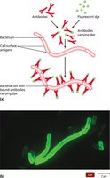
Immunofluorescence
Immunofluorescence uses antibodies conjugated to fluorophores to detect specific molecules in or on cells, aiding in the identification of cell types and proteins.
Application: Used in research and diagnostics to study protein localization and cell identification.
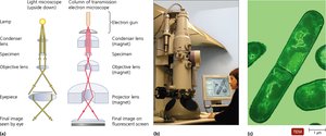
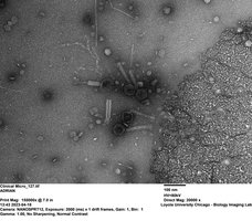
Electron Microscopy
Transmission Electron Microscopy (TEM)
TEM uses electrons instead of light to visualize specimens at very high resolution. Electrons pass through the specimen, and the image is formed based on electron scattering.
Application: Used to study internal structures of cells and viruses.
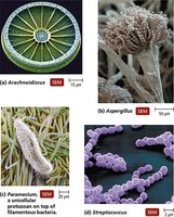
Scanning Electron Microscopy (SEM)
SEM focuses electrons on the surface of a specimen, producing detailed 3D images of its surface structure.
Application: Used to study surface morphology of cells and microorganisms.
Staining Techniques in Microbiology
Types of Staining Procedures
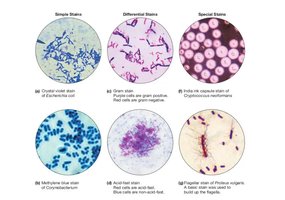
Staining is crucial for visualizing and differentiating microorganisms. There are several types of staining procedures used in microbiology.
Simple staining: Uses a single dye to highlight cell shape, size, and arrangement.
Differential staining: Uses multiple dyes to distinguish between different types of cells or structures (e.g., Gram stain, acid-fast stain).
Special staining: Used for specific structures such as capsules, flagella, and endospores.
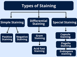
Simple Stains
Simple stains use a single basic dye to color cells, making it easier to observe their morphology.
Common dyes: Crystal violet, methylene blue, safranin
Application: Used to observe Escherichia coli and Staphylococcus aureus
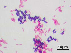
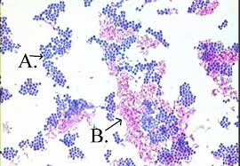
Differential Staining: Gram Stain
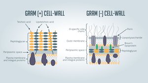
The Gram stain differentiates bacteria based on cell wall structure. Gram-positive bacteria stain purple, while Gram-negative bacteria stain pink.
Gram-positive: Thick peptidoglycan layer retains crystal violet stain.
Gram-negative: Thin peptidoglycan layer and outer membrane; does not retain crystal violet, stains pink with safranin.
Differential Staining: Acid-Fast Stain
Acid-fast staining is used for bacteria with waxy cell walls, such as Mycobacterium. Acid-fast bacteria retain the primary stain even after exposure to acid alcohol.
Application: Diagnosis of mycobacterial infections (e.g., tuberculosis).
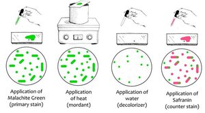
Special Staining: Endospore Stain
Endospore staining differentiates tough, dormant structures formed by bacteria such as Bacillus and Clostridium. Malachite green stains endospores, while safranin stains vegetative cells.
Application: Identification of spore-forming bacteria.
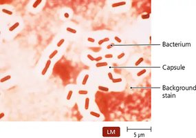
Special Staining: Negative (Capsule) Stain
Negative staining colors the background, leaving capsules visible as clear halos around cells. Capsules protect bacteria from the immune system.
Application: Identification of encapsulated bacteria such as Klebsiella pneumoniae.
Special Staining: Flagella Stain
Flagella stains are used to visualize bacterial flagella, which are too thin to be seen with standard light microscopy. Special stains bind to flagella, making them visible.
Application: Identification of motile bacteria such as Proteus vulgaris.
