 Back
BackChp 4 Microscopy, Staining, and Classification: Study Notes for Microbiology
Study Guide - Smart Notes
 Tailored notes based on your materials, expanded with key definitions, examples, and context.
Chapter 4: Microscopy, Staining, and Classification
Introduction to Microscopy
Microscopy is a fundamental technique in microbiology, enabling scientists to observe microorganisms and cellular structures that are invisible to the naked eye. Understanding the principles of microscopy, including magnification, resolution, and contrast, is essential for accurate observation and classification of microbes.
Units of Measurement in Microscopy
Accurate measurement is crucial in microbiology. Scientists use the metric system for consistency and precision, especially when measuring microscopic entities.
Meter (m): Standard unit of length.
Micrometer (µm): Commonly used to measure bacteria and cells.
Nanometer (nm): Used for viruses and molecular structures.
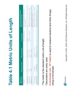
Example: The size of most bacteria ranges from 0.5 to 5 µm, while viruses are typically 20–300 nm.
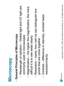
General Principles of Microscopy
Microscopy relies on the interaction of light or electrons with specimens to produce magnified images. Key principles include:
Wavelength of Radiation: Visible light and UV light are commonly used; shorter wavelengths yield higher resolution.
Magnification: The process of enlarging the appearance of an object.
Resolution: The ability to distinguish two points as separate. Higher resolution allows for clearer detail.
Contrast: Differences in intensity between an object and its background; staining and phase techniques enhance contrast.
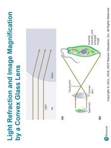
Light Refraction and Magnification
Microscopes use lenses to bend (refract) light, focusing it to magnify specimens. Convex glass lenses are standard in light microscopes.
Refraction: Bending of light as it passes through different media (e.g., air to glass).
Magnification: Determined by the focal length and arrangement of lenses.
Resolution in Microscopy
Resolution is the smallest distance between two objects that can still be distinguished as separate. It is a critical factor in microscopy.
Formula:
Numerical Aperture: A property of the lens affecting resolution.
Shorter wavelengths and higher numerical aperture improve resolution.
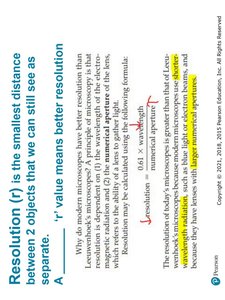
Example: Electron microscopes use electrons (shorter wavelength) for higher resolution than light microscopes.
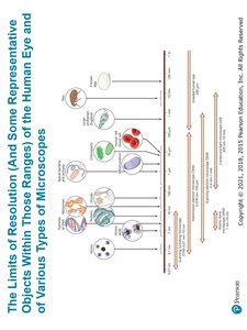
Limits of Resolution and Types of Microscopes
Different microscopes have varying resolving powers, allowing observation of different biological structures.
Human Eye: Can resolve objects down to about 0.2 mm.
Light Microscopes: Resolve objects down to about 0.2 µm.
Electron Microscopes: Resolve objects down to about 0.2 nm.
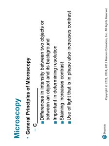
Contrast in Microscopy
Contrast is essential for distinguishing specimens from their background. It is enhanced by staining and phase techniques.
Staining: Increases contrast by coloring cells or structures.
Phase Contrast: Uses differences in light phase to enhance contrast without staining.
Types of Light Microscopy
Light microscopes use visible light to observe specimens. There are several types, each with unique features:
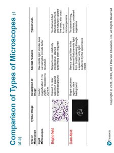
Bright-Field Microscopes: Use direct light; most common in labs.
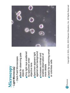
Dark-Field Microscopes: Best for observing pale objects; only light scattered by specimen enters the lens.
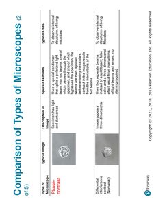
Phase Microscopes: Used for living organisms; contrast is created by differences in light phase.
Differential Interference Contrast Microscopes: Provide 3D appearance.
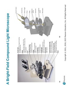
Bright-Field Compound Light Microscope
The compound microscope uses multiple lenses to achieve higher magnification and resolution. It is widely used in microbiology labs.
Objective Lenses: Provide primary magnification.
Ocular Lenses: Further magnify the image.
Total Magnification:
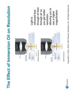
Effect of Immersion Oil on Resolution
Immersion oil is used with high-power objective lenses to reduce light refraction and increase resolution.
Oil Immersion: Light is refracted less through oil than air, allowing higher magnification and better resolution.
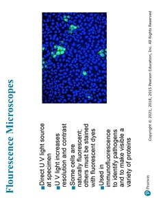
Fluorescence Microscopy
Fluorescence microscopes use UV light to excite fluorescent dyes or proteins, making specific structures visible.
Direct UV Light: Enhances resolution and contrast.
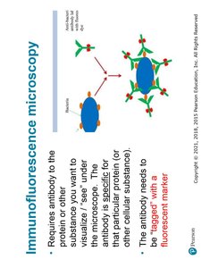
Immunofluorescence: Uses antibodies tagged with fluorescent markers to identify specific proteins or pathogens.

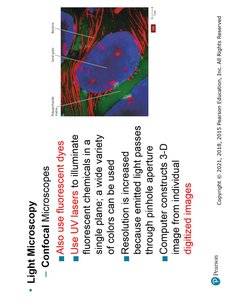
Confocal Microscopy
Confocal microscopes use lasers and fluorescent dyes to produce high-resolution, three-dimensional images of specimens.
UV Lasers: Illuminate chemicals in a single plane.
3D Imaging: Computer constructs 3D images from digitized data.
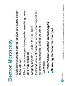
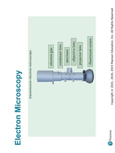
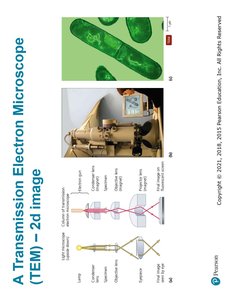
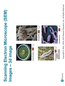
Electron Microscopy
Electron microscopes use beams of electrons instead of light, allowing much higher resolution and magnification.
Transmission Electron Microscope (TEM): Produces 2D images of internal structures.
Scanning Electron Microscope (SEM): Produces 3D images of surface structures.
Magnification: Up to 100,000x or more.
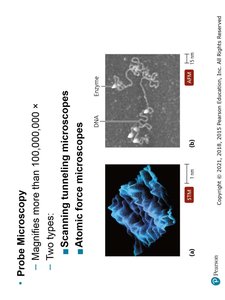
Probe Microscopy
Probe microscopes, such as scanning tunneling and atomic force microscopes, can magnify objects more than 100,000,000x, allowing visualization of molecules and atoms.
Scanning Tunneling Microscopes: Visualize surface at atomic level.
Atomic Force Microscopes: Provide 3D images of surfaces.
Comparison of Types of Microscopes
Different microscopes are suited for different applications in microbiology. The following tables summarize their features:
Type | Special Features | Typical Uses | Image |
|---|---|---|---|
Bright-field | Simple, direct light | General observation | See image_33 |
Dark-field | Scattered light, dark background | Pale objects, motility | See image_33 |
Phase contrast | Enhances contrast without staining | Living cells | See image_34 |
Differential interference contrast | 3D appearance | Detailed cell structure | See image_34 |
Fluorescence | Uses UV light, fluorescent dyes | Immunology, protein localization | See image_35 |
Confocal | UV lasers, 3D imaging | Cell structure, 3D reconstructions | See image_35 |
Transmission Electron | Electrons, 2D images | Internal cell structure | See image_36 |
Scanning Electron | Electrons, 3D images | Surface structure | See image_36 |
Review Questions and Applications
Understanding microscopy is essential for identifying and studying microorganisms. Review questions help reinforce key concepts:
Which is the largest: nanometer, micrometer, or millimeter?
Which unit is most appropriate to measure the size of cells?
The various forms of radiation differ in their wavelength.
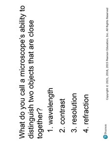
What do you call a microscope's ability to distinguish two objects that are close together? (Resolution)
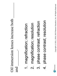
Oil immersion lenses increase both magnification and resolution.
Example: Oil immersion is used in microbiology labs to observe bacteria at high magnification and resolution.
----------------------------------------
