 Back
BackMicroscopy, Staining, and Classification: Study Notes for Microbiology Students
Study Guide - Smart Notes
 Tailored notes based on your materials, expanded with key definitions, examples, and context.
Microscopy, Staining, and Classification
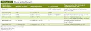
Units of Measurement
Understanding the metric system is essential in microbiology for measuring microorganisms and their structures. The metric system is a decimal-based system used globally, with units for length, mass, volume, and temperature.
Length: Measured in meters (m), with common prefixes such as kilo (k, 1000), centi (c, 1/100), milli (m, 1/1000), micro (µ, 1/1,000,000), and nano (n, 1/1,000,000,000).
Mass: Measured in grams (g).
Volume: Measured in liters (l).
Temperature: Measured in degrees Celsius (°C). Conversion: $\text{Celsius} = \frac{5}{9}(\text{Fahrenheit} - 32)$
Decimal System: Each unit is 1/10th the size of the next unit.
Metric Unit | Meaning of Prefix | Metric Equivalent | U.S. Equivalent | Microbiological Application |
|---|---|---|---|---|
Meter (m) | — | 1 m | 39.37 in | Length of pipet, tape, saline solution prep |
Centimeter (cm) | 1/100 | 0.01 m | 0.39 in | Diameter of bacterial colony |
Millimeter (mm) | 1/1000 | 0.001 m | 0.04 in | Diameter of a bacterial colony |
Micrometer (µm) | 1/1,000,000 | 0.000001 m | 0.00004 in | Diameter of bacterial cells |
Nanometer (nm) | 1/1,000,000,000 | 0.000000001 m | 0.00000004 in | Diameter of poliovirus |
Microscopy
Microscopy is the science of using light or electrons to magnify objects, allowing scientists to observe microorganisms and their structures. The field began with Antoni van Leeuwenhoek and has since evolved to include various types of microscopes.
General Principles of Microscopy
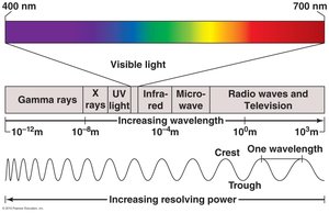
Wavelength of Radiation: The distance between two corresponding parts of a wave. Visible light ranges from 400nm to 700nm.
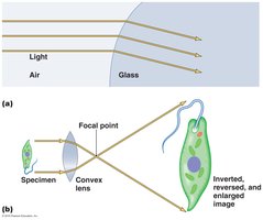
Magnification: The apparent increase in the size of an object, achieved by refracting light through lenses.
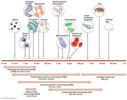
Resolution: The ability to distinguish between objects that are close together. Modern microscopes can resolve objects as small as 0.2 µm.
Contrast: Differences in intensity between objects and their background, often enhanced by staining.
Magnification
Magnification occurs when a beam of light refracts as it passes through a lens, focusing light rays on a focal point. The curvature of the lens determines how light is bent and focused.
Resolution
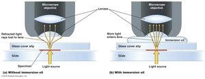
Resolution is critical for distinguishing fine details in specimens. It depends on the wavelength of radiation and the numerical aperture of the lens. Shorter wavelengths (e.g., blue light or electron beams) provide better resolution.
Contrast
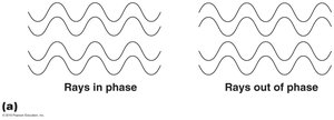
Contrast is essential for visualizing microorganisms, which are often colorless. Staining and using light in phase can increase contrast.
Types of Microscopes
Different microscopes are used depending on the specimen and desired detail.
Light Microscopy
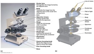
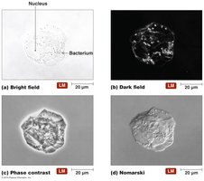
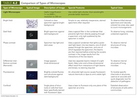
Bright-field Microscopes: Use light to illuminate specimens. Simple microscopes have a single lens; compound microscopes use multiple lenses for higher magnification.
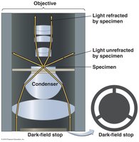
Dark-field Microscopes: Best for observing pale objects; only scattered light enters the objective lens, making specimens appear light against a dark background.
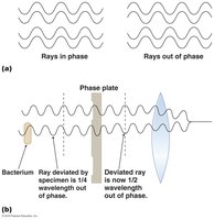
Phase Microscopes: Used for living organisms; create contrast by exploiting differences in phase of light waves.
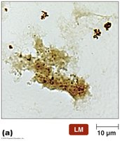
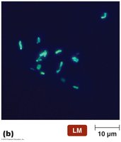
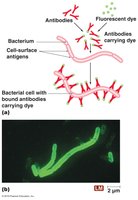
Fluorescence Microscopes: Use UV light to excite fluorescent specimens, increasing resolution and contrast.
Confocal Microscopes: Use UV lasers to create optical slices and construct 3-D images.
Electron Microscopy
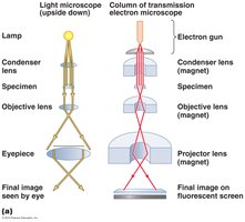
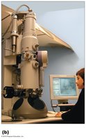
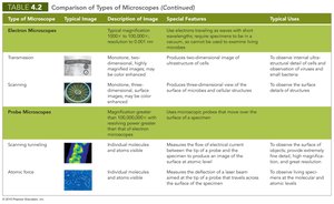
Electron microscopes use electron beams for much higher magnification and resolution than light microscopes. They are essential for viewing viruses, internal cell structures, and molecules.
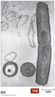
Transmission Electron Microscopes (TEM): Produce 2-D images by passing electrons through specimens.
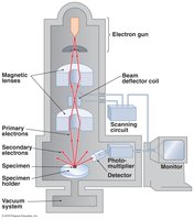
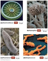
Scanning Electron Microscopes (SEM): Produce 3-D images by scanning the surface of specimens coated with metal.
Probe Microscopy
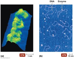
Probe microscopes are advanced instruments that use a physical probe to scan the surface of specimens, achieving magnifications over 100 million times.
Scanning Tunneling Microscopes (STM): Measure electron flow between probe and specimen.
Atomic Force Microscopes (AFM): Use a probe to touch the specimen surface, measuring atomic topography.
Microscope Types Comparison
Microscopes are compared based on their typical images, features, and uses.
Staining
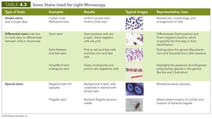
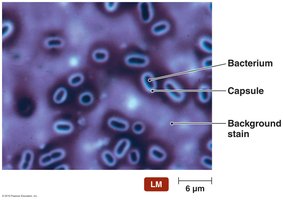
Staining is a technique used to color specimens, increasing contrast and making microorganisms easier to view. Different stains are used for light and electron microscopy.
Smear Preparation: Thin film of organism is spread, air-dried, and fixed (chemical or heat fixation).
Dyes: Usually salts with colored chromophores. Basic dyes stain acidic structures; acidic dyes stain alkaline structures.
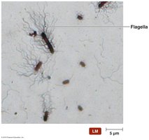
Types of Stains: Simple stains (single dye), differential stains (multiple dyes), special stains (reveal specific structures).
Gram Stain
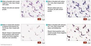
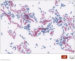
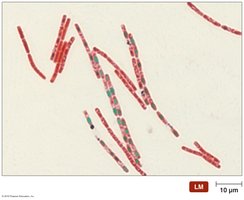
The Gram stain differentiates between Gram-positive (purple) and Gram-negative (pink) bacteria, based on cell wall properties.
Procedure: Crystal violet (primary stain), iodine (mordant), alcohol/acetone (decolorizer), safranin (counterstain).
Applications: Identifies bacterial groups for diagnosis and treatment.
Classification and Identification of Microorganisms
Taxonomy is the science of classifying organisms into groups (taxa) based on similarities. It includes classification, nomenclature, and identification.
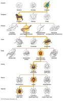
Linnaean System: Groups organisms into species, genera, families, orders, classes, phyla, and kingdoms.
Binomial Nomenclature: Two-part names (Genus species), e.g., Homo sapiens.
Domains: Based on rRNA sequences: Eukarya, Bacteria, Archaea.
Physical Characteristics
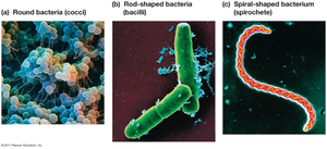
Microorganisms can be identified by their morphology, colony appearance, and cell shape (coccus, bacillus, spiral).
Biochemical Tests
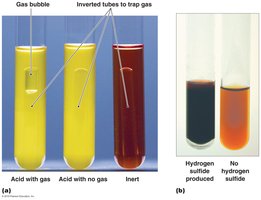
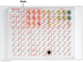
Biochemical tests classify organisms based on their metabolic abilities, such as fermenting carbohydrates or producing waste products.
Serological Tests
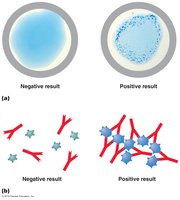
Serological tests detect specific antibodies in blood, using agglutination to identify antigenic microorganisms.
Phage Typing
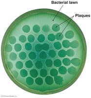
Phage typing identifies bacteria based on their susceptibility to specific bacteriophages, which create plaques where bacteria are killed.
Analysis of Nucleic Acids
Determining the percentage of guanine and cytosine (G+C content) in DNA helps classify prokaryotes.
Taxonomic Keys
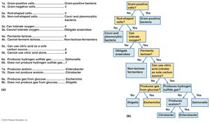
Dichotomous keys are used to identify organisms by guiding users through paired statements to reach a taxonomic identification.
 Additional info: These notes expand on brief points with academic context, definitions, and examples to ensure completeness and clarity for exam preparation.
