 Back
Microscopy, Staining, and Classification: Study Notes for Microbiology
Study Guide - Smart Notes
 Tailored notes based on your materials, expanded with key definitions, examples, and context.
Microscopy, Staining, and Classification
General Principles of Microscopy
Microscopy is fundamental to microbiology, enabling visualization of organisms too small for the naked eye. The effectiveness of a microscope depends on several key principles:
Wavelength of Radiation: The distance between two consecutive crests or troughs in a wave. Shorter wavelengths provide higher resolution, allowing finer details to be observed.
Magnification: The process of enlarging the appearance of an object. It is determined by the product of the magnification of the objective and ocular lenses.
Resolution: The ability to distinguish two points as separate entities. Higher resolution is achieved with shorter wavelengths and proper lens quality.
Contrast: The difference in intensity between an object and its background. Contrast is enhanced by staining and by using light that is in phase.
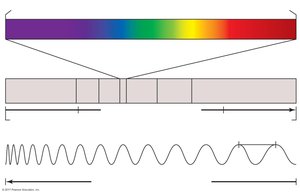
Example: Visible light microscopes use wavelengths between 400–700 nm, while electron microscopes use much shorter wavelengths for greater resolution.
Metric Units of Length in Microbiology
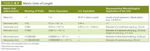
Microbiologists use metric units to measure microorganisms, ranging from meters to nanometers. Understanding these units is essential for interpreting microscopic observations.
Unit | Metric Equivalent | Representative Microbiological Application |
|---|---|---|
Meter (m) | 1 m | Length of pork tapeworm |
Decimeter (dm) | 0.1 m | Diameter of a mushroom cap |
Centimeter (cm) | 0.01 m | Diameter of a bacterial colony |
Millimeter (mm) | 0.001 m | Length of a flea |
Micrometer (µm) | 0.000001 m | Diameter of a typical bacterium |
Nanometer (nm) | 0.000000001 m | Diameter of poliovirus |
Light Refraction and Magnification
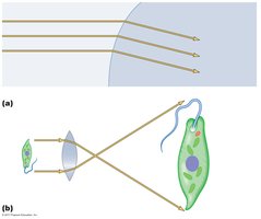
Microscopes use lenses to bend (refract) light, focusing it to produce magnified images. Convex lenses invert, reverse, and enlarge the image of the specimen.
Refraction: The bending of light as it passes through different media (e.g., air, glass).
Focal Point: The point where light rays converge after passing through a lens.
Example: A compound light microscope uses a series of lenses to achieve higher magnification and resolution.
Limits of Resolution and Types of Microscopes
The resolution of a microscope determines the smallest object that can be distinguished. Different types of microscopes have varying resolving powers, suitable for different applications.
Human Eye: Can resolve objects down to about 200 µm.
Light Microscopes: Resolve objects as small as 200 nm.
Electron Microscopes: Resolve objects down to 0.078 nm.
Probe Microscopes: Can visualize individual molecules and atoms.
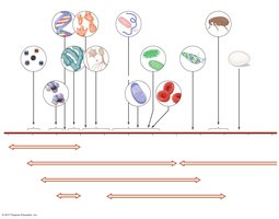
Example: Viruses and proteins require electron or probe microscopes for visualization.
Types of Light Microscopes
Light microscopes are commonly used in microbiology. They include simple and compound bright-field microscopes, as well as specialized types for enhanced contrast.
Simple Microscope: Contains a single magnifying lens, similar to a magnifying glass.
Compound Microscope: Uses a series of lenses for greater magnification. Includes objective and ocular lenses, and often a condenser lens.
Oil Immersion Lens: Increases resolution by reducing light refraction.
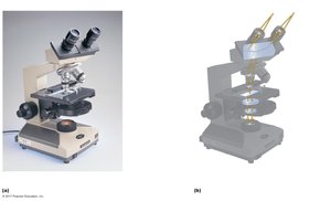
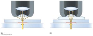
Formula: Total magnification = magnification of objective lens × magnification of ocular lens
Example: Leeuwenhoek used a simple microscope to observe microorganisms.
Comparison of Types of Microscopes
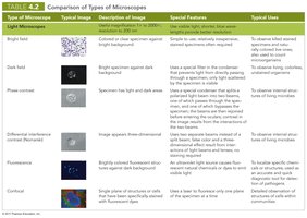
Microscopes are classified based on their imaging principles and applications. The following tables summarize the main types:
Type | Typical Image | Description | Special Features | Typical Uses |
|---|---|---|---|---|
Bright field | Colored or clear specimen | Background is bright | Simple, widely used | Observe stained specimens |
Dark field | Bright specimen, dark background | Special filter for light | Observe living, unstained organisms | |
Phase contrast | Image appears three-dimensional | Phase plate enhances contrast | Observe internal structures | |
Differential interference contrast | Brightly colored, three-dimensional | Uses two beams of light | Observe fine details | |
Fluorescence | Bright specimen, dark background | Uses UV light | Identify specific cells | |
Confocal | Single plane of cells | Uses laser | Detailed imaging of cell layers |
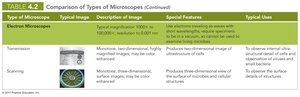
Type | Typical Image | Description | Special Features | Typical Uses |
|---|---|---|---|---|
Transmission Electron Microscope (TEM) | Monochrome, highly magnified | Internal structure of cells | Electrons pass through specimen | Observe internal cell structures |
Scanning Electron Microscope (SEM) | Monochrome, three-dimensional | Surface details | Electrons scan surface | Observe surface details |
Type | Typical Image | Description | Special Features | Typical Uses |
|---|---|---|---|---|
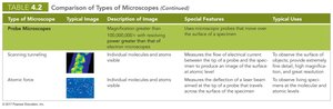
Scanning Tunneling Microscope | Individual molecules and atoms | Measures flow of electrons | Atomic resolution | Observe surface of atoms |
Atomic Force Microscope | Individual molecules and atoms | Measures deflection of laser beam | Atomic resolution | Observe surface of molecules |
Contrast and Staining
Contrast is crucial for distinguishing microorganisms from their background. Staining increases contrast and resolution, making cells and structures more visible.
Staining: Coloring specimens with dyes to enhance visibility.
Preparation: Specimens are spread, air-dried, and fixed before staining.
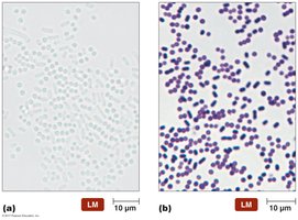
Example: Stained cells are easier to observe and differentiate than unstained cells.
Principles of Staining
Stains are usually salts with a colored portion called the chromophore. Dyes are classified as acidic or basic, depending on their affinity for cellular structures.
Acidic Dyes: Stain alkaline structures.
Basic Dyes: Stain acidic structures; more common since most cells are negatively charged.
Example: Crystal violet, safranin, and methylene blue are basic dyes used in simple stains.
Types of Stains
Stains are categorized as simple, differential, or special, each serving distinct purposes in microbiology.
Simple Stains: Use a single dye to determine cell size, shape, and arrangement.
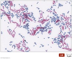
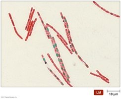
Differential Stains: Use multiple dyes to distinguish between cell types or structures. Examples include Gram stain, acid-fast stain, and endospore stain.
Special Stains: Identify specific structures such as capsules, flagella, or spores.
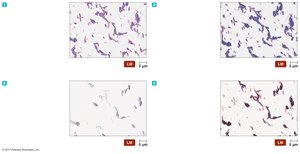
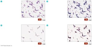
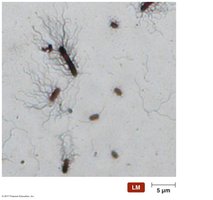
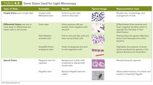
Type of Stain | Examples | Typical Image | Representative Uses |
|---|---|---|---|
Simple Stains | Crystal violet, methylene blue | Uniform purple stain | Reveal cell size, morphology |
Differential Stains | Gram, Ziehl-Neelsen, Schaeffer-Fulton | Gram-positive/negative, acid-fast, endospores | Identify cell types, pathogens |
Special Stains | Negative, flagellar | Background or flagella stained | Identify capsules, flagella |
Classification and Identification of Microorganisms
Taxonomy: Classification, Nomenclature, and Identification
Taxonomy organizes information about organisms, enabling predictions and understanding evolutionary relationships. It includes classification, nomenclature, and identification.
Linnaeus: Developed a system based on common characteristics and binomial nomenclature.
Kingdoms: Originally two, later expanded to five (Animalia, Plantae, Fungi, Protista, Prokaryotae).
Domains: Carl Woese proposed three domains (Eukarya, Bacteria, Archaea) based on rRNA sequences.
Example: Modern taxonomy emphasizes phylogenetic hierarchy and genetic comparisons.
Taxonomic and Identifying Characteristics
Microorganisms are identified using physical, biochemical, serological, and genetic characteristics.
Physical Characteristics: Morphology of protozoa, fungi, algae, and worms; colony appearance.
Biochemical Tests: Ability to utilize or produce chemicals; used to identify pathogens.
Serological Tests: Study of antigen-antibody reactions; antibodies identify organisms.
Phage Typing: Use of bacteriophages to identify bacteria.
Analysis of Nucleic Acids: Genetic sequencing for classification.
Example: Biochemical tests distinguish prokaryotes by their metabolic capabilities.
Dichotomous Keys
Dichotomous keys are tools for identifying organisms based on a series of paired statements. Each choice directs the user to another pair or provides the organism's name.
Structure: Series of "either/or" statements.
Application: Used in laboratory settings for identification of bacteria and other microorganisms.
Example: A dichotomous key may distinguish Gram-positive from Gram-negative bacteria, then further classify based on shape, oxygen tolerance, and biochemical properties.
