 Back
BackLecture Notes Chapter 4 and 5
Study Guide - Smart Notes
 Tailored notes based on your materials, expanded with key definitions, examples, and context.
Microscopy, Staining, and Classification
Types of Light Microscopy
Microscopy is essential for visualizing microorganisms, which are too small to be seen with the naked eye. Different types of light microscopy provide unique advantages for observing cell structure, motility, and specific cellular components.
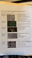
Microscope Type | Image | Main Features |
|---|---|---|
Brightfield |
| Light passes directly through the specimen. Most common for stained, fixed samples. Provides good contrast for stained cells but poor for live, unstained cells. |
Darkfield | Only light scattered by the specimen enters the objective lens, making specimens appear bright against a dark background. Useful for observing live, unstained organisms and motility. | |
Fluorescence | Uses fluorescent dyes or proteins to label specific cell structures. Cells or structures appear brightly colored against a dark background. Allows for high specificity in identifying cellular components. | |
Phase-Contrast | Enhances contrast in transparent specimens without staining. Useful for observing live cells and their internal structures. | |
Electron Microscopy | Uses electron beams instead of light, providing much higher resolution. Transmission EM (TEM) shows internal structures; Scanning EM (SEM) shows surface details. |
Staining Techniques
Staining increases contrast and allows for differentiation of cellular components. Common stains include:
Simple Stain: Uses a single dye to highlight the entire organism.
Gram Stain: Differentiates bacteria into Gram-positive (purple) and Gram-negative (pink) based on cell wall structure.
Acid-Fast Stain: Identifies Mycobacterium species by staining waxy cell walls.
Endospore Stain: Highlights bacterial endospores, which are resistant to harsh conditions.
Cell Structure and Function
Bacterial Cell Wall and Morphology
Bacterial cells have diverse shapes and structural features that are critical for classification and function. The cell wall provides shape, protection, and is a key target for antibiotics.
Coccus: Spherical shape.
Bacillus: Rod-shaped.
Spirillum: Spiral-shaped.
Vibrio: Comma-shaped.
Spirochete: Flexible, spiral-shaped.
Gram-positive bacteria have thick peptidoglycan layers, while Gram-negative bacteria have thin peptidoglycan and an outer membrane.
Bacterial Motility
Bacteria move using structures such as flagella, pili, or by gliding. Motility is important for colonization and infection.
Flagella: Long, whip-like structures for movement.
Pili: Short, hair-like structures for attachment and sometimes movement.
Axial Filaments: Found in spirochetes, enabling corkscrew motion.
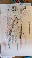
Microbial Metabolism
Enzyme Function and Kinetics
Enzymes are biological catalysts that speed up chemical reactions in cells. They lower the activation energy required for reactions and are highly specific for their substrates.
Active Site: The region on the enzyme where the substrate binds.
Enzyme-Substrate Complex: Temporary association between enzyme and substrate during the reaction.
Product Formation: Substrate is converted into product, which is then released.

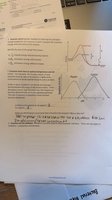
The rate of enzyme-catalyzed reactions can be described by the Michaelis-Menten equation:
v: Reaction rate
Vmax: Maximum rate
[S]: Substrate concentration
Km: Substrate concentration at half-maximal velocity
Enzyme Inhibition
Enzyme activity can be regulated by inhibitors:
Competitive Inhibitors: Bind to the active site, blocking substrate binding.
Noncompetitive Inhibitors: Bind to another part of the enzyme, changing its shape and reducing activity.
Microbial Growth and Classification
Microbial Growth Phases
Bacterial populations grow in distinct phases when cultured in a closed system:
Lag Phase: Cells adapt to new environment; little to no division.
Log (Exponential) Phase: Rapid cell division and population growth.
Stationary Phase: Growth rate slows as resources become limited; cell death balances division.
Death Phase: Cells die at an exponential rate due to depletion of nutrients and accumulation of waste.
Classification of Microorganisms
Microorganisms are classified based on morphology, staining characteristics, metabolic properties, and genetic analysis. The Gram stain is a primary method for bacterial classification.
Gram-Positive: Thick peptidoglycan, stains purple.
Gram-Negative: Thin peptidoglycan, outer membrane, stains pink.
Summary Table: Microscopy and Staining Methods
Method | Purpose | Key Features |
|---|---|---|
Brightfield | General observation of stained cells | Good for fixed, stained samples; poor for live cells |
Darkfield | Observation of live, unstained cells | Bright specimens on dark background |
Fluorescence | Specific labeling of cell structures | Uses fluorescent dyes; high specificity |
Phase-Contrast | Live cell imaging | Enhances contrast without staining |
Electron Microscopy | High-resolution imaging | Visualizes ultrastructure; TEM and SEM types |
Additional info: These notes integrate foundational concepts from microscopy, cell structure, and microbial metabolism, as well as practical laboratory techniques for observing and classifying microorganisms. The diagrams and tables reinforce key distinctions and processes relevant to microbiology students.
