 Back
BackMicroscopy: Structure, Function, and Proper Use of the Compound Light Microscope
Study Guide - Smart Notes
 Tailored notes based on your materials, expanded with key definitions, examples, and context.
Microscopy
Parts of the Compound Light Microscope
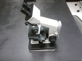
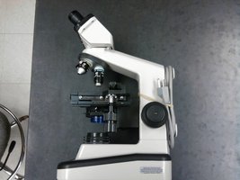
The compound light microscope is an essential tool in microbiology, allowing for the visualization of microorganisms and cellular structures. Understanding its components and their functions is crucial for accurate observation and safe handling.
Arm: The supporting structure used to carry the microscope.
Base: The bottom platform that provides stability.
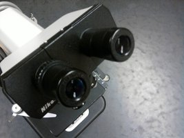
Ocular (Eyepiece): The lens through which the specimen is viewed, typically with a magnification of 10x.
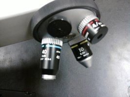
Nosepiece: The rotating part that holds multiple objective lenses.
Objective Lenses: Lenses of varying magnification (commonly 4x, 10x, 40x, and 100x) used to view specimens at different levels of detail.
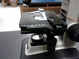
Stage: The platform where the slide is placed for observation.
Condenser: Focuses light onto the specimen.
Focus Knobs:
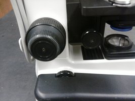
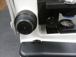
Coarse Focus Knob: Moves the stage up and down significantly for initial focusing.
Fine Focus Knob: Allows for precise focusing adjustments.
Stage Motion Knobs: Move the stage horizontally and vertically to position the slide.
Lamp: Provides the light source necessary for viewing specimens.
Magnification and Optical Path
Microscopes use a combination of lenses to magnify specimens. The total magnification is calculated by multiplying the magnification of the ocular lens by that of the objective lens.
Ocular Magnification: Usually 10x.
Objective Lens Magnifications: Commonly 4x, 10x, 40x, and 100x.
Total Magnification:
4x objective × 10x ocular = 40x
10x objective × 10x ocular = 100x
40x objective × 10x ocular = 400x
100x objective × 10x ocular = 1000x
Light Path: Light from the lamp passes through the condenser, then through the specimen, objective lens, and finally the ocular lens.
Proper Handling and Common Mistakes
Correct handling of the microscope is essential to prevent damage and ensure accurate results. Several common mistakes should be avoided:
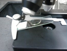
Never place a slide under the stage clip: This can crack the slide.
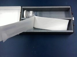
Never clean the lens with paper towels: This will scratch the lens. Always use lens paper.
Never use the coarse adjustment knob with the high power (40x or 100x) objective lens: This can crack both the slide and the lens. Use only the fine adjustment knob at high magnification.
Stage and Slide Handling
The stage is the platform where the slide rests. Proper placement and movement of the slide are crucial for observation and safety.
Stage Motion Knobs: Allow precise movement of the slide for scanning.
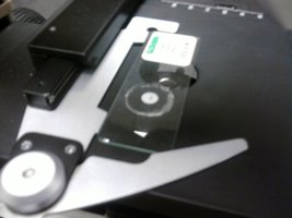
Stage Clips: Secure the slide in place.
Correct Slide Placement: Place the slide gently on the stage and secure it with the clips, avoiding forceful placement.
Summary Table: Microscope Parts and Functions
Part | Function |
|---|---|
Arm | Support and carrying |
Base | Stability |
Ocular | Viewing, magnifies 10x |
Nosepiece | Holds objectives |
Objective Lenses | Magnify specimen (4x, 10x, 40x, 100x) |
Stage | Holds slide |
Condenser | Focuses light |
Focus Knobs | Coarse/fine focusing |
Stage Motion Knobs | Moves slide |
Lamp | Light source |
Lab Quiz Information
Students should be prepared for a lab quiz covering the microscopy labs. Practice quizzes are available for extra credit and review.
Quiz Format: Ten questions, 1.5 points each.
Practice Quiz: Available online, extra credit, unlimited attempts.
Preparation: Review all microscope parts, functions, and proper handling procedures.
Additional info:
Microscopy is foundational for studying microorganisms, enabling visualization of cells, bacteria, and other microbes.
Proper care and handling of the microscope ensure longevity and accuracy in laboratory observations.
