 Back
BackMicroscopy Techniques for Observing Living Microorganisms
Study Guide - Smart Notes
 Tailored notes based on your materials, expanded with key definitions, examples, and context.
Examination of Living Microorganisms
Introduction to Observing Living Microbes
Microscopy is a fundamental technique in microbiology for examining the structure, motility, and behavior of living microorganisms. Two common methods for observing live microbes are the wet mount and hanging drop techniques, each with specific advantages and limitations.
Microscopy Techniques
Brightfield Microscopy
Brightfield microscopy is a standard light microscopy technique where the specimen appears dark against a bright background.
It is commonly used to observe unstained microorganisms in wet mounts and hanging drop slides.
Provides basic visualization but may lack contrast for transparent organisms.
Wet Mount Technique
The wet mount technique involves suspending a specimen in a liquid on a microscope slide, covering it with a coverslip, and observing it under a microscope. This method is simple and allows for the examination of living, unstained microorganisms in their natural state.
Advantages: Quick preparation, observation of live movement, and natural morphology.
Limitations: Low contrast, rapid evaporation, and potential compression of organisms by the coverslip.
Wet Mount Procedure
Stir or shake the infusion to suspend organisms.
Transfer a drop to a slide using a Pasteur pipette or inoculating loop.
Carefully place a coverslip over the drop and gently press to spread the liquid.
Observe under low and high power objectives, adjusting light as needed.
For motility studies, alcohol or Gram’s iodine may be used to reduce movement.
Hanging Drop Technique
The hanging drop method is used to observe live microorganisms in a three-dimensional, hydrated environment, minimizing evaporation and preventing compression. This technique is especially useful for studying microbial motility.
Advantages: Preserves natural motility, reduces evaporation, and prevents crushing of delicate organisms.
Limitations: More complex preparation, not suitable for oil immersion objectives.
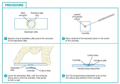
Hanging Drop Procedure
Apply a ring of petroleum jelly around the concavity of a depression slide.
Place a loopful of microbial suspension on a coverslip.
Invert the depression slide over the coverslip so the drop is suspended in the well.
Examine under low or medium power objectives.
Comparison: Wet Mount vs. Hanging Drop
Wet Mount: Easier and faster to prepare, but organisms may be compressed and dry out quickly.
Hanging Drop: Maintains hydration, prevents compression, and allows for extended observation of motility.
Motility in Microorganisms
Brownian Movement vs. True Motility
Brownian Movement: Random, erratic motion of particles due to collisions with molecules in a fluid. Not a form of true motility.
True Motility: Directed movement powered by cellular structures such as flagella or cilia.
Examples of Brownian Movement
Pollen grains in water
Dust particles in air
Ink particles dispersing in water
Staining and Chemical Effects
Use of Alcohol and Gram’s Iodine
Alcohol: Disrupts cell membranes, leading to cell death or reduced motility.
Gram’s Iodine: Denatures proteins and enzymes, inactivating flagella and disrupting cellular function.
Microbiomes
Definition and Composition
A microbiome is a community of microorganisms, including protozoa, algae, fungi, and bacteria, that inhabit a particular environment. Larger members of the microbiome can be observed using the hanging drop technique.
Microscopy and Microbial Size
Size Ranges and Magnification
Organism | Approximate Size | Brightfield Magnification | Lab Notes |
|---|---|---|---|
Viruses | ~20–300 nm | Not visible | Require electron microscopy |
Bacteria | ~0.5–5 µm | 1000× (oil immersion) | Usually require staining (Gram stain) |
Fungi (Yeast) | ~3–10 µm | 400×–1000× | Often show budding |
Fungi (Molds/Hyphae) | Width ~5–10 µm | 100×–400× | Branching structures easily seen |
Protozoa | ~10–100 µm | 100×–400× | Often motile; may be unstained |
Algae | ~10 µm to several mm | 40×–400× | Larger species visible at low power |
Prokaryotes vs. Eukaryotes
Key Differences
Feature | Prokaryotes | Eukaryotes |
|---|---|---|
Nucleus | No true nucleus; DNA in nucleoid region | Membrane-bound nucleus |
Organelles | No membrane-bound organelles | Contains membrane-bound organelles |
Size | 0.1 to 5.0 µm | 10 to 100 µm |
Genetic Material | Single, circular DNA; may have plasmids | Multiple, linear chromosomes; no plasmids (typically) |
Ribosomes | 70S (smaller) | 80S (larger) |
Cell Division | Binary fission | Mitosis and meiosis |
Examples | Bacteria, Archaea | Plants, Animals, Fungi, Protists |
Bacterial Infections
Common Sites and Causative Agents
Bacterial infections can affect various body systems, with specific organisms commonly associated with each site.
Site | Common Pathogens |
|---|---|
Skin Infections | Staphylococcus aureus, Streptococcus pyogenes, Pseudomonas aeruginosa |
Sexually Transmitted Diseases | Chlamydia trachomatis, Neisseria gonorrhoeae, Treponema pallidum, Ureaplasma urealyticum, Haemophilus ducreyi |
Urinary Tract Infections | Escherichia coli, Other Enterobacteriaceae, Staphylococcus saprophyticus, Pseudomonas aeruginosa |
Frequently Asked Questions
Why should the drop be hanging in the hanging drop method?
Prevents compression of organisms, allowing for natural movement.
Minimizes evaporation, maintaining hydration for longer observation.
Provides a clear, three-dimensional observation field.
Why is oil immersion not used with the hanging drop procedure?
Oil immersion could disrupt or contaminate the suspended drop, causing collapse or altered behavior.
Lower-power objectives (10x or 40x) are sufficient for observing motility in the hanging drop method.
Why are microorganisms hard to see in wet preparations?
Many microorganisms are nearly transparent and blend into the liquid background.
Rapid movement of live organisms makes them difficult to focus and observe in detail.
