 Back
BackMini-Textbook Study Notes: Introduction to Prokaryotic Cells
Study Guide - Smart Notes
 Tailored notes based on your materials, expanded with key definitions, examples, and context.
Introduction to Prokaryotic Cells
Overview of Prokaryotic Life
Prokaryotic cells are the most ancient and smallest life forms on Earth, inhabiting the planet for billions of years. They are classified into two domains: Archaea and Bacteria. These cells are unicellular, lack a membrane-bound nucleus, and have a simpler genetic makeup compared to eukaryotic cells. Prokaryotes thrive in diverse environments due to their varied extracellular and intracellular structures.
Domains: Archaea and Bacteria
Key Features: Unicellular, lack nucleus, lack membrane-bound organelles
Importance: Understanding prokaryotes aids in disease prevention, treatment, and drug development.
Prokaryotic Cell Basics
Domains and Classification
Prokaryotic cells are divided into two domains: Bacteria and Archaea. While both are prokaryotes, genetic studies show that Archaea are more closely related to Eukarya than to Bacteria.

Similarity: Both lack a nucleus and membrane-bound organelles.
Difference: Archaea have unique membrane lipids and cell wall components compared to Bacteria.
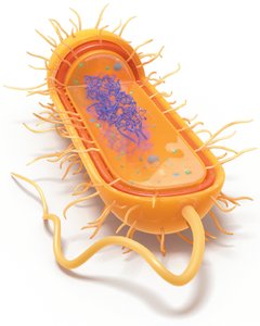
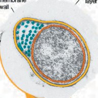
Prokaryotic Cell Structure
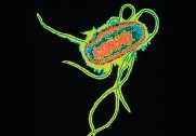
Prokaryotic cells have a variety of structures, including a plasma membrane, cell wall, cytoplasm, ribosomes, and sometimes external features like capsules, fimbriae, and flagella.
Size, Shape, and Arrangement
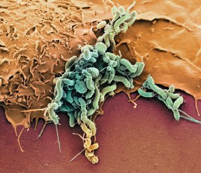
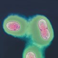
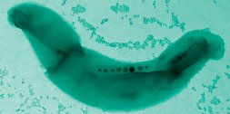
Prokaryotes exhibit diverse shapes and arrangements, which are important for identification and survival. Most prokaryotes are between 0.2 and 2.0 μm in diameter. Their small size allows efficient nutrient diffusion.
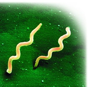
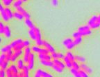
Shapes: Cocci (spherical), Bacilli (rod-shaped), Vibrio (comma-shaped), Spirochete (spiral), Stella (star-shaped), Filamentous, Pleomorphic (variable shape)
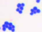
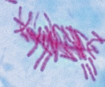
Arrangements: Single, Diplo (pairs), Strep (chains), Staph (clusters), Palisades (side-by-side rods)
Binary Fission
Most prokaryotes reproduce asexually by binary fission, a process that results in two genetically identical daughter cells. The steps include DNA replication, cell elongation, septum formation, partitioning, and separation.
Key Point: Binary fission is simpler than mitosis and does not introduce genetic variation.

Extracellular Structures
Plasma Membrane Structure and Function
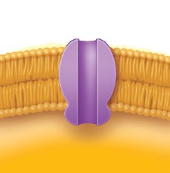
The plasma membrane is a phospholipid bilayer that acts as a selective barrier and a site for metabolic reactions. It is described by the fluid-mosaic model, where lipids and proteins move laterally within the bilayer.
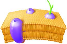
Bacterial Membranes: Linear fatty acids, ester linkages
Archaeal Membranes: Branched isoprenoids, ether linkages, can form monolayers

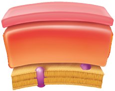
Cell Wall Structure and Function
The cell wall provides rigidity and protection. Bacterial cell walls contain peptidoglycan, while archaeal cell walls may contain pseudopeptidoglycan or other polymers. The Gram stain differentiates bacteria based on cell wall structure:
Gram-positive: Thick peptidoglycan, teichoic acids, stains purple
Gram-negative: Thin peptidoglycan, outer membrane with lipopolysaccharide (LPS), stains pink
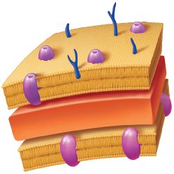
Table: Comparison of Gram-Positive and Gram-Negative Bacteria
Feature | Gram-Negative | Gram-Positive |
|---|---|---|
Outer membrane | Yes | No |
Lipid A (endotoxin) | Yes | No |
Porins | Yes | No |
Teichoic acids | No | Yes |
Peptidoglycan | Thin | Thick |
Gram stain color | Pink | Purple |
Resistance to drying | No | Yes |
Penicillin susceptibility | Low | High |
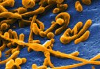
Acid-Fast Bacteria
Acid-fast bacteria, such as Mycobacterium and Nocardia, have waxy mycolic acid in their cell walls, making them resistant to decolorization by acid-alcohol and slow-growing. Acid-fast staining is important for diagnosing diseases like tuberculosis and leprosy.
Mycoplasma and L-Forms
Mycoplasma species lack a cell wall and are pleomorphic. L-forms are bacteria that lose their cell wall due to mutation or stress. Both are resistant to antibiotics targeting cell wall synthesis.
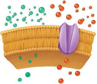
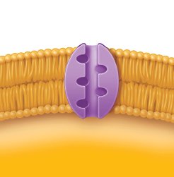
Transport Mechanisms
Prokaryotic cells use passive and active transport to move substances across their membranes:
Passive Transport: Simple diffusion, facilitated diffusion, osmosis (no energy required)
Active Transport: Requires energy (ATP or ion gradients); includes primary active transport, secondary active transport (symport/antiport), and phosphotransferase systems

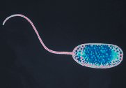
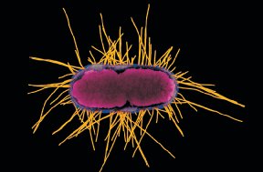
Motility and Adhesion Structures
Flagella: Provide motility; arrangements include monotrichous, lophotrichous, amphitrichous, and peritrichous.
Periplasmic Flagella: Located between the plasma membrane and cell wall, enabling corkscrew motion in spirochetes.
Fimbriae: Short, bristle-like structures for adhesion.
Pili: Longer, less numerous structures for adhesion, motility, and gene transfer.
Glycocalyx: Carbohydrate-rich layer for protection and adhesion; can be a slime layer (loose) or capsule (tight).


Intracellular Structures
Nucleoid
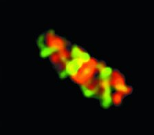
The nucleoid is the region where the prokaryotic chromosome (usually a single, circular DNA molecule) is located. It is not membrane-bound.

Ribosomes
Prokaryotic ribosomes are 70S, composed of 50S and 30S subunits. They are the site of protein synthesis and are structurally distinct from eukaryotic ribosomes, supporting the endosymbiotic theory.

Cytoskeleton
The prokaryotic cytoskeleton is made of protein filaments that provide structure, aid in cell division, and help maintain cell shape. These proteins are functionally similar to eukaryotic actin and tubulin.
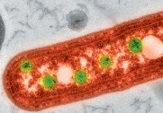
Inclusion Bodies
Inclusion bodies are storage sites for nutrients and other substances, such as glycogen, poly-β-hydroxybutyrate (PHB), and magnetosomes. They help cells survive in nutrient-poor conditions and may play roles in metabolism and navigation.
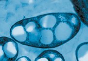
Endospores
Endospores are metabolically inactive, highly resistant structures formed by certain bacteria (notably Bacillus and Clostridium) to survive harsh conditions. They can withstand extreme heat, drying, chemicals, and radiation. Endospores are not reproductive structures; one cell forms one spore.
Sporulation: Formation of endospore under stress
Germination: Return to vegetative state when conditions improve
Clinical Importance: Endospores are difficult to eradicate and require special disinfection protocols in healthcare settings.

Summary Table: Key Prokaryotic Structures and Functions
Structure | Function |
|---|---|
Cell wall | Protection, shape, prevents lysis |
Plasma membrane | Selective barrier, metabolic site |
Nucleoid | Genetic material (DNA) |
Ribosome | Protein synthesis |
Cytoskeleton | Shape, division, organization |
Inclusion bodies | Storage of nutrients |
Flagella | Motility |
Fimbriae/Pili | Adhesion, gene transfer (pili) |
Glycocalyx | Protection, adhesion |
Endospore | Dormancy, survival in harsh conditions |
Key Concepts for Exam Preparation
Prokaryotes are classified into Archaea and Bacteria, both lacking a nucleus and membrane-bound organelles.
Cell wall structure is central to Gram staining and clinical identification.
Prokaryotic cells use various transport mechanisms to move substances across membranes.
Motility and adhesion are mediated by flagella, fimbriae, pili, and glycocalyx.
Endospores are highly resistant structures formed by some bacteria for survival.
