 Back
Negative Staining in Microbiology: Principles, Procedure, and Interpretation
Study Guide - Smart Notes
 Tailored notes based on your materials, expanded with key definitions, examples, and context.
Negative Staining: Principles and Practice
Background and Chemical Principles
Negative staining is a microbiological technique used to visualize cell morphology without distorting cellular structures. It employs acidic dyes with an anionic (negatively charged) chromophore. Because bacterial cells are also negatively charged, the dye is repelled, resulting in a stained background while the cells remain clear and visible.
Acidic dyes: Examples include Nigrosine and Congo red.
Basic dyes: Have a positively charged chromophore and stain the cell directly.
Negative stain: Reveals basic morphological information (shape, arrangement, size) but not internal structures.
No fixation required: Unlike simple stains, negative staining does not require heat or chemical fixation, preserving cell morphology.
Example: The negative stain is ideal for observing delicate structures such as capsules or for accurate measurement of cell size.
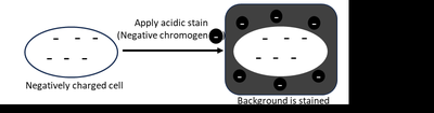
Comparison: Simple Stain vs Negative Stain
Simple stain: Uses a basic dye to stain the cell directly; requires fixation, which may distort cell shape.
Negative stain: Uses an acidic dye to stain the background; does not require fixation, preserving true cell morphology.
Table: Comparison of Staining Methods
Staining Method | Dye Type | Fixation Required | Stains | Reveals |
|---|---|---|---|---|
Simple Stain | Basic (positive chromophore) | Yes | Cell | Shape, arrangement, size |
Negative Stain | Acidic (negative chromophore) | No | Background | Shape, arrangement, size |
Procedure for Negative Staining
The negative staining procedure is designed to minimize distortion and maximize clarity of cell morphology. The steps are as follows:
Add dye: Place a drop of nigrosine on one end of a clean slide.
Add specimen: Transfer bacteria into the drop of dye using an inoculation loop or sterile toothpick (for oral samples).
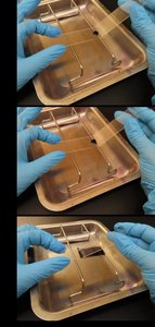
Spread: Use a second slide at a 30˚ angle to spread the mixture across the slide in one fluid motion.
Air dry: Allow the slide to air dry completely (5–10 minutes). Do not heat-fix.
Dispose: Place the spreader slide in disinfectant.
Observe: Examine the slide under oil immersion after drying.
Interpretation of Negative Stain Results
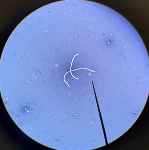
After staining, cells appear clear against a dark background. This allows for accurate assessment of cell shape, arrangement, and size. For example, Bacillus subtilis and Staphylococcus epidermidis can be distinguished based on their morphology and arrangement.
Cell morphology: Shape (cocci, bacilli, spirilla, etc.)
Arrangement: Chains, clusters, pairs, etc.
Size: Measured accurately due to lack of distortion
Advantages and Limitations of Negative Staining
Advantages:
Preserves true cell morphology (no distortion from fixation)
Quick and simple procedure
Useful for observing capsules and delicate structures
Limitations:
Does not reveal internal cell structures
Cannot differentiate cell wall characteristics
Lab Report and Questions
Students are expected to record observations regarding specimen morphology, arrangement, and magnification. Key questions include:
Range of bacterial morphologies observed
Comparison of cheek cells to bacterial cells
True/False: Negative staining requires two or more dyes; requires a fixed smear; more accurately reveals cell morphology than simple staining
Identification: Cell arrangement, shape, size (but not cell wall characteristics)
Properties: Acidic, stains background, negative charge, contains chromogen
Sketch: Appearance of Staphylococcus aureus stained with nigrosine
Advantage: More accurate morphology due to lack of fixation
Key Terms and Definitions
Acidic dye: Dye with a negatively charged chromophore, stains background.
Basic dye: Dye with a positively charged chromophore, stains cell.
Chromophore: The colored ion in a dye responsible for staining.
Fixation: Process of adhering cells to a slide, often distorts morphology.
Cell morphology: Shape and arrangement of cells.
Additional info: Negative staining is especially useful for visualizing bacterial capsules, which are not easily stained by conventional methods. The technique is also used in clinical microbiology for rapid assessment of sample quality and cell types.
