 Back
BackObserving Microorganisms Through a Microscope: Principles and Techniques
Study Guide - Smart Notes
 Tailored notes based on your materials, expanded with key definitions, examples, and context.
Microscopes and Magnification
Introduction to Microscopy
Microscopy is essential in microbiology for visualizing organisms too small to be seen with the naked eye. Microscopes use lenses to magnify specimens, allowing for detailed observation of microbial structure and function.
Magnification: The process of enlarging the appearance of an object.
Resolution: The ability to distinguish two points as separate entities; higher resolution reveals finer details.
Microscopy Range: Different microscopes have varying resolving powers, allowing observation of objects from millimeters to nanometers in size.
Key Concept: Resolution increases with decreasing wavelength of the illuminating source.
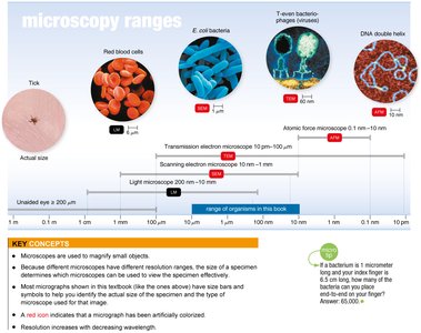
Types of Microscopes
Simple and Compound Microscopes
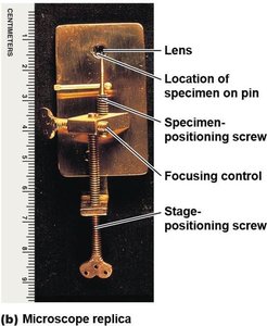
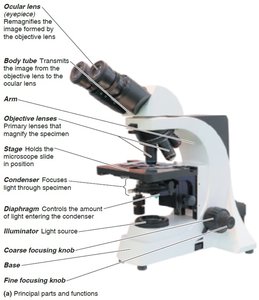
Simple microscopes use a single lens, while compound microscopes use multiple lenses for higher magnification and resolution. The total magnification is the product of the objective and ocular lens magnifications.
Simple Microscope: Similar to a magnifying glass but with a higher quality lens.
Compound Microscope: Utilizes both objective and ocular lenses for enhanced magnification.
Total Magnification Formula:
Light Microscopy
Principles and Types
Light microscopy uses visible light to illuminate specimens. Several specialized techniques enhance contrast and detail for different applications.
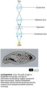
Brightfield Microscopy: Produces a dark image against a bright background; most common for stained specimens.
Darkfield Microscopy: Illuminates specimens against a dark background, useful for observing live, unstained organisms.
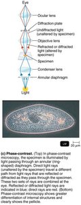
Phase-Contrast Microscopy: Enhances contrast in transparent specimens by amplifying differences in refractive index; ideal for viewing living cells and internal structures.
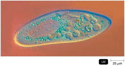
Differential Interference Contrast (DIC) Microscopy: Uses prisms to produce high-contrast, pseudo-3D images with color differences.
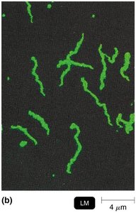
Fluorescence Microscopy: Uses UV light to excite fluorescent dyes, causing specimens to emit visible light; useful for identifying specific microbes.
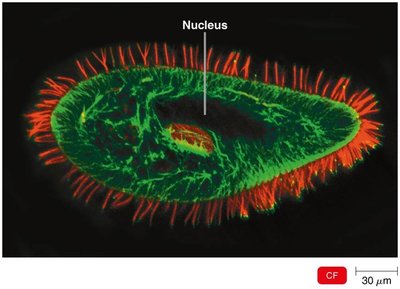
Confocal Microscopy: Uses lasers and fluorescent dyes to create sharp, three-dimensional images of thick specimens.
Electron Microscopy
Transmission and Scanning Electron Microscopes
Electron microscopes use electron beams instead of light, providing much higher resolution and magnification. They are essential for visualizing viruses and subcellular structures.
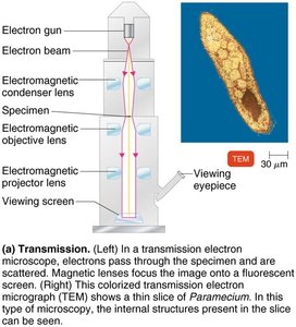
Transmission Electron Microscopy (TEM): Electrons pass through ultrathin specimen sections, revealing internal structures in high detail.
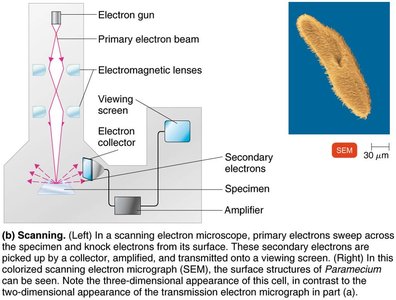
Scanning Electron Microscopy (SEM): Electrons scan the specimen surface, producing detailed three-dimensional images of surface structures.
Resolution: TEM can resolve structures as small as 10 pm; SEM typically resolves down to 10 nm.
Staining Techniques in Microbiology
Preparing Smears and Staining
Staining enhances contrast and allows visualization of microbial morphology and structure. Smears are thin films of microorganisms fixed to slides before staining.
Simple Stain: Uses a single basic dye to highlight the entire microorganism.
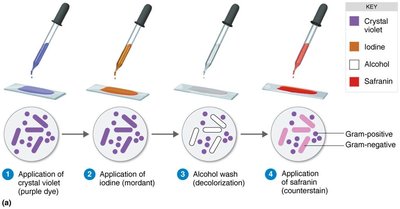
Differential Stains: Distinguish between different types of bacteria based on cell wall properties.
Gram Stain: Differentiates bacteria into Gram-positive (thick peptidoglycan) and Gram-negative (thin peptidoglycan, outer membrane) groups.
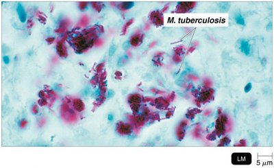
Acid-Fast Stain: Identifies bacteria with waxy cell walls (e.g., Mycobacterium), which retain dye after acid-alcohol decolorization.
Summary Table: Acid-Fast Staining Results
Step | Acid-Fast Bacteria | Non-Acid-Fast Bacteria |
|---|---|---|
Primary Stain: Carbolfuchsin | Red | Red |
Decolorizing Agent: Acid-Alcohol | Red | Colorless |
Counterstain: Methylene Blue | Red | Blue |
Key Terms and Concepts
Magnification: Enlargement of an object's image.
Resolution: Ability to distinguish two points as separate.
Refractive Index: Measure of light-bending ability of a medium.
Chromophore: Colored ion in a stain.
Mordant: Substance used to fix or intensify a stain.
