 Back
Observing Microorganisms Through a Microscope: Principles and Applications
Study Guide - Smart Notes
 Tailored notes based on your materials, expanded with key definitions, examples, and context.
Observing Microorganisms Through a Microscope
Introduction to Microscopes
Microorganisms are typically too small to be seen with the naked eye and require the use of microscopes for visualization. Microscopes are essential tools in microbiology, allowing scientists to study the structure, function, and behavior of cells and microorganisms.
Microscope: An optical instrument used to visualize objects too small to be seen by the unaided eye, such as cells and microbes.
There are two main types of microscopes commonly used in microbiology:
Light Microscopes: Use visible light to magnify small objects, typically up to 1000X.
Electron Microscopes: Use beams of electrons for much higher magnification and resolution, allowing visualization of structures at the molecular level.
The range of objects visible to the human eye, light microscopes, and electron microscopes varies significantly.
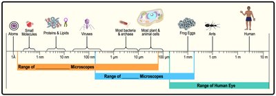
Types of Microscopes and Their Applications
Different types of microscopes are suited for different applications in microbiology, depending on the size and nature of the specimen.
Light microscopes are suitable for viewing most bacteria, archaea, and eukaryotic cells.
Electron microscopes are required for visualizing viruses, proteins, and smaller molecules.
Practice Example: To visualize prokaryotic cells, both light and electron microscopes can be used, but for muscle proteins, an electron microscope is preferred due to the small size of proteins.
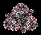
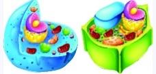
Light Microscopy
Introduction to Light Microscopy
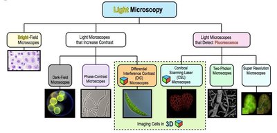
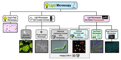
Light microscopy uses visible light and a series of lenses to magnify objects up to about 1000X. There are several types of light microscopes, each with unique advantages for visualizing different specimens.
Major classes of light microscopes include:
Bright-field Microscopes
Microscopes that increase contrast (e.g., phase-contrast, dark-field, differential interference contrast)
Microscopes that detect fluorescence
Bright-Field Microscopy
The bright-field microscope is the most common type of light microscope, generating a bright background against which specimens appear darker. It is routinely used to examine both stained and unstained specimens.
Drawback: Unstained, transparent organisms have low contrast and are difficult to see.
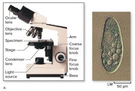
Compound Light Microscope: Uses multiple lenses (objective and ocular) for increased magnification.
Total Magnification: Calculated as the product of the magnification of the ocular lens and the objective lens.
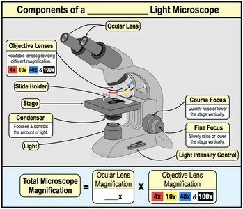
Formula for Total Magnification:
Components of the Compound Light Microscope
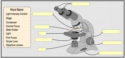
The compound light microscope consists of several key components, each with a specific function:
Ocular Lens (Eyepiece): Magnifies the image produced by the objective lens.
Objective Lenses: Provide different levels of magnification (e.g., 4x, 10x, 40x, 100x).
Stage: Holds the specimen slide.
Condenser Lens: Focuses and controls the amount of light reaching the specimen.
Coarse and Fine Focus Knobs: Used to bring the specimen into sharp focus.
Light Source: Illuminates the specimen.
Light Microscopes That Increase Contrast
Specialized light microscopes are designed to increase contrast, making it easier to visualize unstained, transparent cells. These include:
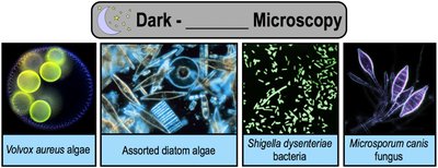
Dark-Field Microscopes: Observe bright specimens against a dark background by directing light at an angle.
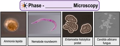
Phase-Contrast Microscopes: Enhance contrast by amplifying differences in refractive index, making dense structures appear darker.
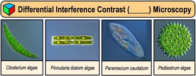
Differential Interference Contrast (DIC) Microscopes: Produce highly detailed, 3D-like images with contrasting colors.
Dark-Field Microscopy
Uses a special condenser to allow only light scattered by the specimen to reach the objective lens, causing the specimen to appear bright against a dark background.
Phase-Contrast Microscopy
Makes cells and their dense structures appear darker than the gray background, ideal for viewing living, unstained cells.
Differential Interference Contrast (DIC) Microscopy
Creates detailed, high-contrast, 3D-like images of live specimens, often with added color for contrast.
Light Microscopes That Detect Fluorescence
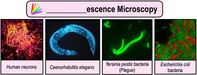
Fluorescence microscopy uses the ability of certain molecules to absorb short wavelengths of light and emit longer wavelengths, making fluorescent objects stand out against a dark background.
Fluorescence Microscopes: Project ultraviolet light onto the specimen, causing it to fluoresce. Fluorochromes can be used to tag specific molecules or structures.
Immunofluorescence: Combines fluorochromes with antibodies to tag specific targets within cells.
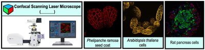
Confocal Scanning Laser Microscopes (CSLMs)
Use lasers and computer control to generate high-contrast, 3D images, allowing visualization of different planes within a specimen.
Two-Photon Microscopes (TPM)
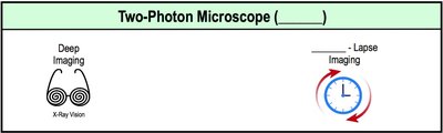
Use two photons and less damaging light for deep imaging and time-lapse studies of living cells.
Super-Resolution Microscopes (SRM)
Achieve resolutions as fine as 0.01 μm, allowing visualization of molecules that are too close together to be seen as distinct with conventional light microscopes.
Electron Microscopy
Introduction to Electron Microscopes
Electron microscopes use beams of electrons instead of light, providing much higher magnification (up to 10,000,000X) and resolution (~0.3 nm). They are essential for studying structures at the molecular and atomic levels.
Specimens must be prepared for viewing in a vacuum and are typically non-living.
Images are black-and-white but can be colorized with computers for clarity.
Transmission Electron Microscope (TEM)
Forms 2D images by passing electrons through thin slices of a specimen, ideal for visualizing internal cell structures.
Drawback: Complex preparation can distort cells and create artifacts.
Scanning Electron Microscope (SEM)
Forms 3D images by detecting electrons scattered from the surface of a specimen, ideal for visualizing surface structures.
Staining Techniques in Microscopy
Introduction to Staining
Many microbes are colorless and transparent, making them difficult to visualize. Staining with dyes increases contrast and allows for better visualization under the microscope.
Different staining techniques are used depending on the purpose of the observation.
Simple Staining
Simple staining uses a single dye to color cells, increasing contrast. There are two main types of dyes:
Basic Dyes: Positively charged, stain the inside of cells by binding to negatively charged cell components.
Acidic Dyes: Negatively charged, stain the background by being repelled by negatively charged cell components (negative staining).
Gram Staining
Gram staining is a differential staining technique that separates bacteria into two groups based on cell wall structure:
Gram-positive bacteria: Stain purple.
Gram-negative bacteria: Stain pink or red.
Steps: Primary stain (crystal violet), iodine (mordant), decolorizer (alcohol), counterstain (safranin).
Special Staining Techniques
Special stains are used to visualize specific structures within or outside cells, such as capsules, endospores, and flagella.
Capsule Stain: Stains the background, making the capsule visible.
Endospore Stain: Identifies bacterial endospores.
Flagella Stain: Visualizes bacterial flagella.
Acid-Fast Stain: Identifies bacteria with mycolic acid in their cell walls (e.g., Mycobacterium).
Fluorescent Dyes and Immunofluorescence
Fluorescent dyes and immunofluorescence techniques are used to tag and visualize specific cell components or molecules. Antibodies conjugated with fluorochromes can specifically bind to antigens, allowing for targeted visualization.
Summary Table: Types of Microscopes and Their Functions
Microscope Type | Main Function |
|---|---|
Bright-Field Microscope | Views stained or unstained specimens on a bright background |
Dark-Field Microscope | Views bright specimens against a dark background |
Phase-Contrast Microscope | Enhances contrast of dense structures in living cells |
Differential Interference Contrast (DIC) Microscope | Produces detailed, high-contrast, 3D-like images |
Fluorescence Microscope | Detects fluorescence from tagged molecules |
Confocal Scanning Laser Microscope | Generates high-contrast, 3D images at different planes |
Two-Photon Microscope | Deep imaging and time-lapse imaging of living cells |
Super-Resolution Microscope | Visualizes molecules at extremely high resolution |
Transmission Electron Microscope (TEM) | 2D images of internal cell structures |
Scanning Electron Microscope (SEM) | 3D images of cell surface structures |
