 Back
Observing Microorganisms Through a Microscope: Principles and Techniques
Study Guide - Smart Notes
 Tailored notes based on your materials, expanded with key definitions, examples, and context.
Ch. 3 Observing Microorganisms Through a Microscope
Microscopes and Magnification
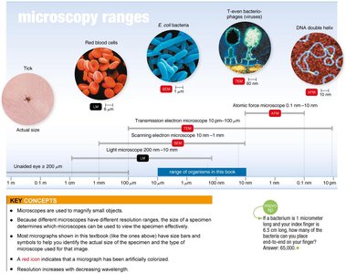
Microscopes are essential tools in microbiology, allowing scientists to observe organisms and structures too small to be seen with the naked eye. The range of microscopes includes light microscopes, electron microscopes, and atomic force microscopes, each with different resolution capabilities.
Magnification: The process of enlarging the appearance of an object.
Resolution: The ability to distinguish two points as separate entities; higher resolution allows for clearer, more detailed images.
Range: Light microscopes can resolve objects from 200 nm to 1 mm, while electron microscopes can resolve down to 0.1 nm.
Microscopy applications: Used to study bacteria, viruses, cell structures, and molecular complexes.
Types of Light Microscopy
Light microscopy utilizes visible light to observe specimens. Compound light microscopes are the most common, and several specialized techniques enhance contrast and detail.
Brightfield Microscopy: Produces a bright background; specimens appear darker.
Darkfield Microscopy: Produces a dark background; specimens appear lighter.
Phase-Contrast Microscopy: Enhances contrast in transparent specimens by combining direct and diffracted light rays.
Principal Parts and Functions of the Compound Light Microscope
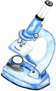
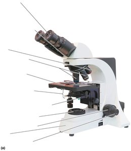
The compound light microscope consists of several key components, each with a specific function in magnifying and resolving specimens.
Ocular lens (eyepiece): Remagnifies the image formed by the objective lens.
Objective lenses: Primary lenses that magnify the specimen.
Stage: Holds the microscope slide in position.
Condenser: Focuses light through the specimen.
Diaphragm: Controls the amount of light entering the condenser.
Illuminator: Light source.
Coarse and fine focusing knobs: Adjust focus.
Body tube: Transmits the image from the objective lens to the ocular lens.
Path of Light in Compound Light Microscopy
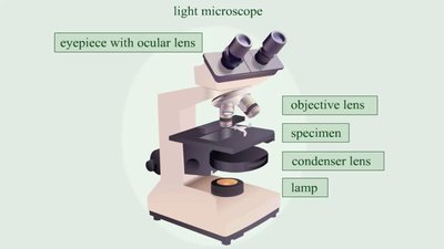
Light passes through the illuminator, condenser, specimen, objective lens, and ocular lens, forming a magnified image for observation.
Total magnification: Calculated as the product of the magnification of the objective lens and the ocular lens.
Resolution: Shorter wavelengths of light provide greater resolution.
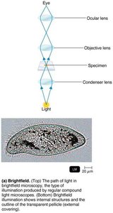
Brightfield, Darkfield, and Phase-Contrast Microscopy
Different illumination techniques provide varying levels of contrast and detail for observing microorganisms.
Brightfield: Internal structures and outlines are visible against a bright background.
Darkfield: Only light reflected by the specimen enters the objective lens, making the specimen appear bright against a dark background.
Phase-Contrast: Combines direct and diffracted light rays to enhance contrast in living cells and internal structures.
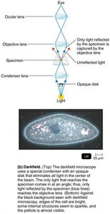
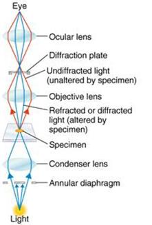
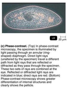
Refraction and Oil Immersion
Refraction is the bending of light as it passes through different media. Immersion oil is used with high-power objective lenses to reduce refraction and increase resolution.
Refractive index: A measure of the light-bending ability of a medium.
Oil immersion: Prevents loss of light due to refraction, improving image clarity.
Preparing Microbes for Staining
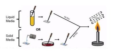
Staining is essential for visualizing microorganisms under the microscope. The process involves preparing a smear, fixing the specimen, and applying stains to highlight cellular structures.
Smear: A thin film of microorganisms spread over a slide.
Fixation: Attaching microorganisms to the slide using heat, which kills them.
Staining: Coloring microorganisms with dyes to emphasize certain structures.
Types of Stains
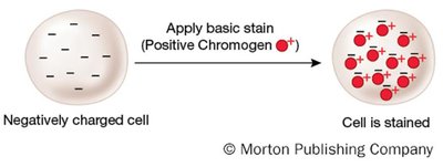
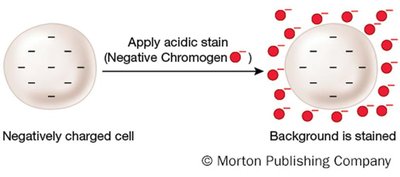
Stains are classified based on the charge of their chromophore and their interaction with the cell.
Basic dye: Chromophore is a cation; stains the cell.
Acidic dye: Chromophore is an anion; stains the background (negative staining).
Simple Stains
Simple stains use a single basic dye to highlight the entire microorganism, making cell shapes and structures visible. A mordant may be used to intensify the stain.
Mordant: A fixative that increases the affinity of the stain for the specimen or thickens structures for better visibility.

Differential Stains
Differential stains react differently with various types of bacteria, allowing for their classification. The most common differential stains are the Gram stain and the acid-fast stain.
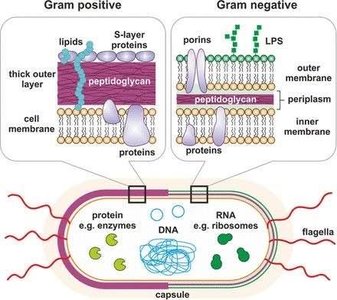
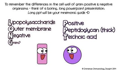
Gram stain: Distinguishes between Gram-positive and Gram-negative bacteria based on cell wall structure.
Acid-fast stain: Identifies bacteria with waxy cell walls, such as Mycobacterium and Nocardia.
Gram Staining Procedure
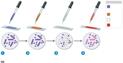
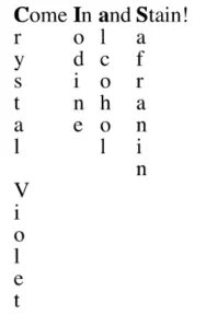
The Gram stain involves four steps: application of crystal violet, iodine (mordant), alcohol (decolorization), and safranin (counterstain).
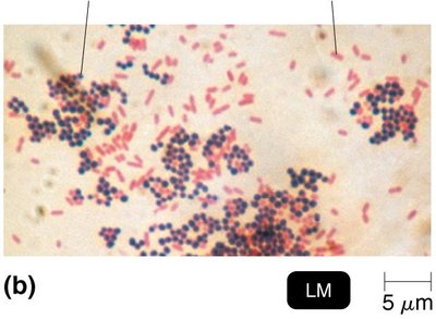
Gram-positive bacteria: Retain crystal violet and appear purple.
Gram-negative bacteria: Lose crystal violet after alcohol wash and take up safranin, appearing pink/red.
Acid-Fast Staining
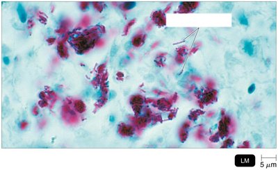
Acid-fast staining is used for bacteria with waxy cell walls that resist decolorization by acid-alcohol. The procedure uses carbolfuchsin as the primary stain, acid-alcohol as the decolorizer, and methylene blue as the counterstain.
Acid-fast bacteria: Retain red color after acid-alcohol treatment.
Non–acid-fast bacteria: Lose red color and take up blue counterstain.
Special Stains
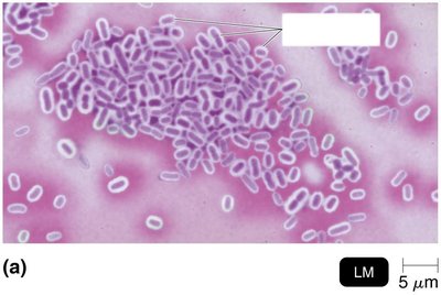
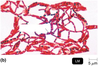
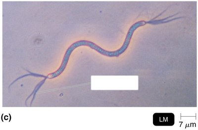
Special stains are used to highlight specific structures within microorganisms, such as capsules, endospores, and flagella.
Capsule stain: Negative staining with India ink or nigrosin; capsules appear as halos.
Endospore stain: Uses malachite green (with heat) and safranin; endospores appear green within red/pink cells.
Flagella stain: Uses a mordant and carbolfuchsin to thicken flagella for visibility.
Summary Table: Staining Techniques
Stain Type | Purpose | Key Steps | Result |
|---|---|---|---|
Simple Stain | Visualize cell shape/structure | Single basic dye | Entire cell colored |
Gram Stain | Differentiate Gram-positive/negative | Crystal violet, iodine, alcohol, safranin | Purple (positive), Pink (negative) |
Acid-Fast Stain | Identify acid-fast bacteria | Carbolfuchsin, acid-alcohol, methylene blue | Red (acid-fast), Blue (non–acid-fast) |
Capsule Stain | Visualize capsules | Negative staining | Capsule appears as halo |
Endospore Stain | Visualize endospores | Malachite green (heat), safranin | Green spores in red cells |
Flagella Stain | Visualize flagella | Mordant, carbolfuchsin | Thickened, visible flagella |
Key Equations
Total Magnification:
Resolution:
Additional info: Academic context was added to clarify the principles of microscopy, staining, and the interpretation of images and tables. The notes are structured to provide a comprehensive overview suitable for exam preparation in a college-level microbiology course.
