Back
Observing Microorganisms through a Microscope: Principles and Techniques
Study Guide - Smart Notes
 Tailored notes based on your materials, expanded with key definitions, examples, and context.
Microscopes in Microbiology
Introduction to Microscopy
Microscopy is essential in microbiology for visualizing microorganisms that are too small to be seen with the naked eye. Various types of microscopes and techniques are used to observe the structure, morphology, and behavior of microbes.
Principles of Light Microscopy
Lenses and Refraction
- Lenses focus light rays at a specific place called the focal point. - The focal length is the distance between the center of the lens and the focal point. - The strength of a lens is inversely related to its focal length: shorter focal length means higher magnification. - Refraction is the bending or change in the angle of light rays as they pass through a medium such as a lens. 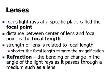
Refractive Index and Light Bending
- Different media (e.g., air and water) have different refractive indices, causing light to bend when transitioning between them. - This principle is important in microscopy, as it affects how light passes through specimens and lenses. 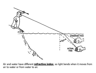
Oil Immersion Technique
- At high magnifications, light may bend in air and miss the objective lens, reducing image clarity. - Immersion oil is used to match the refractive index of glass, minimizing light bending and improving resolution. 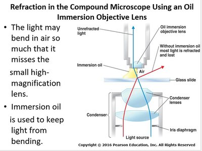
Microscope Resolution and Numerical Aperture
Resolution (Resolving Power)
- Resolution is the ability of a lens to distinguish small objects that are close together. - It is defined as the shortest distance between two points that can still be distinguished as separate. - Wavelength of light is a major factor: shorter wavelengths provide greater resolution. - Numerical aperture (NA) is a measure of a lens's ability to gather light and resolve fine specimen detail at a fixed distance. - Resolving power formula: - Example: Leeuwenhoek’s microscope had a resolution of 1 μm. 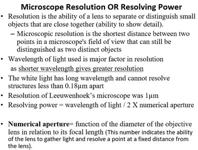
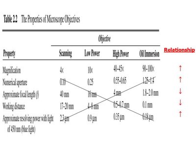
Properties of Microscope Objectives
Property | Scanning | Low Power | High Power | Oil Immersion |
|---|---|---|---|---|
Magnification | 4x | 10x | 40–45x | 90–100x |
Numerical Aperture | 0.10 | 0.25 | 0.55–0.65 | 1.25–1.4 |
Approximate Focal Length (mm) | 40 | 16 | 4.0–4.7 | 1.8–2.0 |
Working Distance (mm) | 17–20 | 4–8 | 0.5–1.0 | 0.16 |
Approximate Resolving Power (blue light, 450 nm) | 2.3 μm | 0.9 μm | 0.53 μm | 0.43 μm |
Types of Light Microscopy
Bright Field vs. Dark Field Microscopy
- Bright field microscopy uses transmitted light to illuminate specimens, which appear dark against a bright background. - Dark field microscopy uses an opaque disk to block direct light, so only light reflected by the specimen enters the objective lens. Specimens appear bright against a dark background. 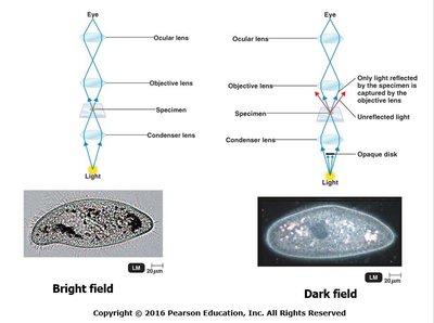
Darkfield Microscopy
- Used to study living microorganisms, especially those that are thin or difficult to stain. - An opaque disc in the condenser blocks direct light; only scattered light from the specimen is observed. - Useful for observing spirochetes such as Treponema pallidum (causes syphilis). 
Electron Microscopy
Transmission Electron Microscope (TEM)
- Electrons pass through a thin specimen and are scattered; magnetic lenses focus the image onto a screen or photographic plate. - Reveals internal structures of cells. - Example: Thin slices of Paramecium can be observed for internal details. 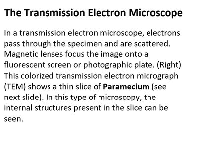
Scanning Electron Microscope (SEM)
- Electrons scan the surface of a specimen, producing images of external structures. - No sectioning is required; the electron beam removes electrons from the specimen’s surface, which are then detected to form an image. 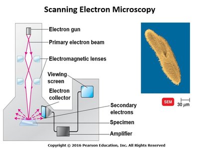
Comparison of TEM and SEM
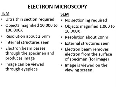
Feature | TEM | SEM |
|---|---|---|
Sectioning | Ultra thin section required | No sectioning required |
Magnification | 10,000–100,000X | 1,000–10,000X |
Resolution | ~2.5 nm | ~20 nm |
Structures Seen | Internal | External |
Image Viewing | Eyepiece | Viewing screen |
Microbial Staining Techniques
Smear Preparation
- A bacterial smear is a dried preparation of bacterial cells on a glass slide. - A good smear is evenly spread, not washed away during staining, and not distorted. - Only a small amount of culture is needed; thick smears hinder light passage and visualization. 
Chemistry of a Stain
- Stains are organic compounds with three parts: 1. Benzene: Organic color solvent 2. Chromophore: Colored chemical group 3. Auxochrome: Ionizes the chromogen to bind with cells, fibers, or tissues - Chromogen = Benzene + Chromophore (colored compound, not a stain by itself) 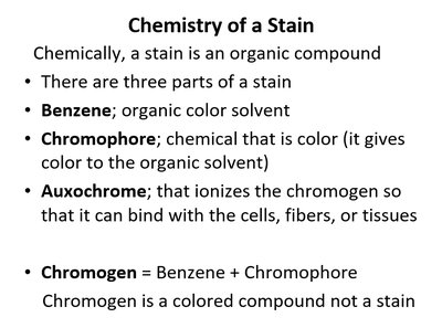
Staining Procedures
- Stains are salts with a colored ion (chromophore): - Basic dyes (chromophore is a cation): e.g., crystal violet, methylene blue - Acidic dyes (chromophore is an anion): e.g., eosin, acid fuchsin - Bacteria are slightly negatively charged, so acidic dyes stain the background (negative staining). - Types of staining procedures: 1. Simple 2. Differential 3. Special 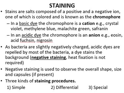
Differential Staining
- Uses at least three chemical reagents applied sequentially to a heat-fixed smear: 1. Primary stain: Colors all cells 2. Decolorizing agent: Removes primary stain from some cells/components 3. Counterstain: Contrasts with the primary stain - After decolorization, cells that retain the primary stain appear one color; others take up the counterstain. 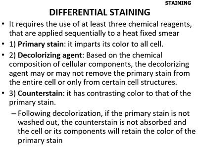
Gram Staining
- Developed by Hans Christian Gram in 1884, this differential staining procedure classifies bacteria as Gram-positive or Gram-negative. - Steps: 1. Apply primary stain (crystal violet) to heat-fixed smear. 2. Add iodine (mordant) to form a crystal violet-iodine complex. 3. Wash with alcohol or acetone (decolorizer). 4. Apply counterstain (usually safranin). - Gram-positive bacteria retain the crystal violet and appear purple; Gram-negative bacteria lose the primary stain and take up the counterstain, appearing pink/red.