 Back
BackObserving Microorganisms Through a Microscope: Principles and Techniques
Study Guide - Smart Notes
 Tailored notes based on your materials, expanded with key definitions, examples, and context.
Observing Microorganisms Through a Microscope
Introduction to Microscopy in Microbiology
Microscopy is fundamental to microbiology, enabling the visualization and study of microorganisms that are otherwise invisible to the naked eye. This chapter covers the principles, types, and applications of various microscopes, as well as staining techniques essential for observing microbial structure and function.
Units of Measurement in Microbiology
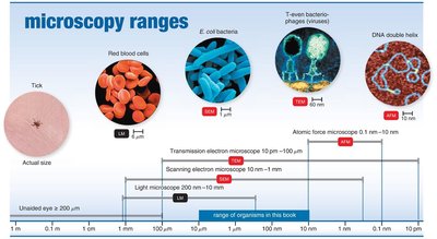
Micrometers and Nanometers
Microorganisms are typically measured in micrometers (µm) and nanometers (nm).
1 µm = 10-6 meters; 1 nm = 10-9 meters.
1000 nm = 1 µm; 0.001 µm = 1 nm.
Example: A typical bacterium is about 1–10 µm in length, while viruses are usually 20–300 nm.
Microscopy: The Instruments
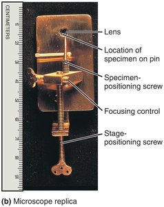
Simple and Compound Microscopes
Simple microscope: Contains a single lens, similar to a magnifying glass but with higher quality.
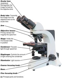
Compound microscope: Uses multiple lenses (objective and ocular) to achieve higher magnification and resolution.
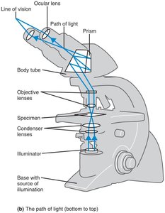
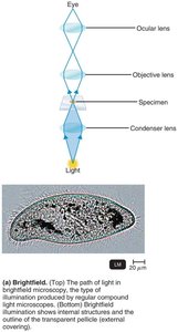
Path of Light in a Compound Microscope
Light passes from the illuminator through the condenser, specimen, objective lens, body tube, and finally the ocular lens.
Total magnification is calculated as:
Resolution and Refractive Index
Resolution (resolving power): The ability to distinguish two points as separate; higher resolution allows for finer detail.
Shorter wavelengths of light provide greater resolution.
Limit of resolution for a compound light microscope is about 0.2 µm, with a maximum useful magnification of ~1500x.
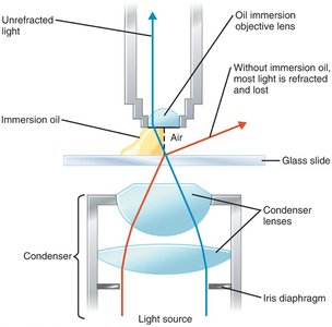
Refractive index: A measure of how much a substance bends light. Immersion oil is used to reduce light refraction and increase resolution at high magnification.
Types of Light Microscopy
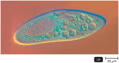
Brightfield Microscopy
Brightfield microscopy is the standard form of light microscopy, where dark objects are visible against a bright background. It is best for stained specimens but may lack contrast for live, unstained cells.
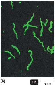
Darkfield Microscopy
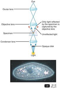
Darkfield microscopy uses an opaque disk to block direct light, so only light reflected by the specimen enters the objective lens. This technique is useful for observing live, unstained microorganisms, such as spirochetes.
Phase-Contrast Microscopy
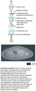
Phase-contrast microscopy enhances contrast in transparent specimens without staining, allowing detailed examination of living cells and internal structures by combining direct and diffracted light rays.
Differential Interference Contrast (DIC) Microscopy
DIC microscopy uses two beams of light and prisms to produce high-contrast, brightly colored, three-dimensional images of live specimens.
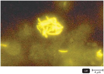
Fluorescence Microscopy
Fluorescence microscopy uses ultraviolet (UV) light to excite fluorescent dyes (fluorochromes) that emit visible light. It is widely used for rapid detection of specific microbes using fluorescent-antibody techniques (immunofluorescence).
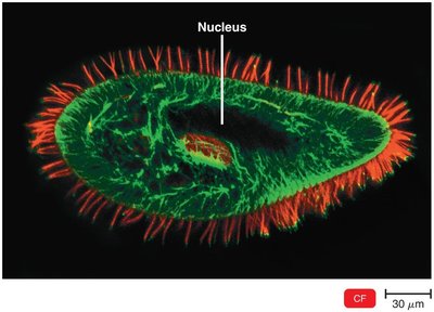
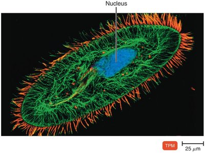
Confocal and Two-Photon Microscopy
Confocal microscopy: Uses lasers and fluorochromes to obtain sharp, two-dimensional images at various depths, which can be reconstructed into three-dimensional images.
Two-photon microscopy: Uses two photons of long-wavelength light to excite dyes, allowing imaging of living cells up to 1 mm deep and tracking cell activity in real time.
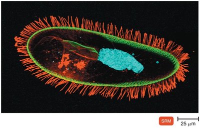
Super-Resolution Light Microscopy
Super-resolution microscopy uses two laser beams to achieve resolution beyond the diffraction limit of light, allowing visualization of structures at the nanometer scale.
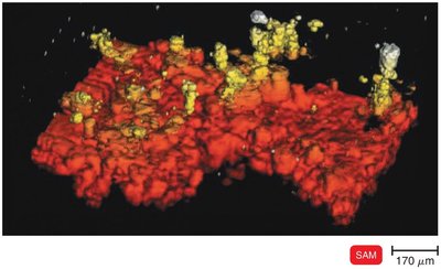
Scanning Acoustic Microscopy (SAM)
SAM measures sound waves reflected from a specimen, useful for studying cells attached to surfaces, such as biofilms, with a resolution of about 1 µm.
Electron Microscopy
Principles of Electron Microscopy
Uses electrons instead of light, providing much greater resolution due to the shorter wavelength of electrons.
Essential for visualizing viruses and internal cellular structures.
Images are black and white but can be digitally colored.
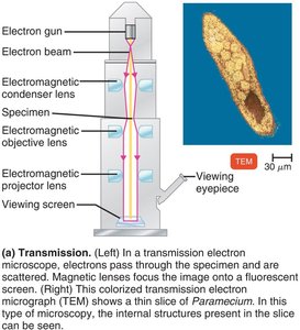
Transmission Electron Microscopy (TEM)
Electrons pass through ultrathin sections of a specimen, revealing internal structures.
Magnification: 10,000–10,000,000x; resolution: 0.2 nm.
Specimens require extensive preparation and are viewed under high vacuum.
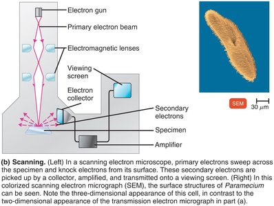
Scanning Electron Microscopy (SEM)
Electrons scan the surface of a specimen, producing detailed three-dimensional images of surface structures.
Magnification: 1,000–500,000x; resolution: 0.5 nm.
Scanned-Probe Microscopy
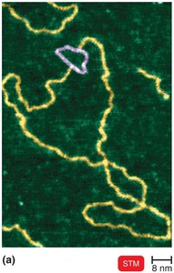
Scanning Tunneling Microscopy (STM)
Uses a tungsten probe to scan the surface at atomic resolution; no special specimen preparation is needed.
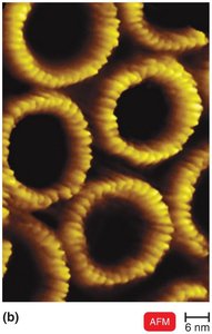
Atomic Force Microscopy (AFM)
Uses a metal-and-diamond probe to scan the specimen, producing three-dimensional images at near-atomic detail.
Preparation of Specimens for Light Microscopy
Staining and Fixation
Staining: Coloring microorganisms with dyes to emphasize structures.
Smear: A thin film of microorganisms spread on a slide.
Fixation: Attaches and kills microorganisms, preserving their structure. Methods include heat or chemical fixation (e.g., methanol).
Types of Dyes
Basic dyes: Chromophore is a cation (e.g., crystal violet, methylene blue, safranin); stains bacterial cells (which are negatively charged).
Acidic dyes: Chromophore is an anion (e.g., eosin, acid fuchsin, nigrosin); used for negative staining (stains background, not cells).
Simple Stains
Use a single basic dye to highlight the entire microorganism, revealing cell shape and structure.
A mordant may be used to intensify the stain or enlarge structures.
Differential Stains
Gram Stain
The Gram stain is a key differential staining technique that classifies bacteria as gram-positive or gram-negative based on cell wall structure.
Step | Gram-Positive | Gram-Negative |
|---|---|---|
Primary Stain: Crystal Violet | Purple | Purple |
Mordant: Iodine | Purple | Purple |
Decolorizer: Alcohol/Acetone | Purple | Colorless |
Counterstain: Safranin | Purple | Pink/Red |
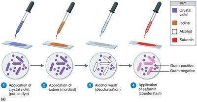
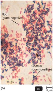
Applications: The Gram stain is essential in clinical microbiology for rapid identification and guiding treatment decisions.
Acid-Fast Stain
The acid-fast stain identifies bacteria with waxy cell walls (e.g., Mycobacterium, Nocardia) that retain the primary stain even after acid-alcohol decolorization.
Step | Acid-Fast | Non–Acid-Fast |
|---|---|---|
Primary Stain: Carbolfuchsin | Red | Red |
Decolorizer: Acid-Alcohol | Red | Colorless |
Counterstain: Methylene Blue | Red | Blue |
Special Stains
Capsule Stain
Capsules are visualized using negative staining (e.g., India ink or nigrosin) followed by a simple stain for the cell.
Capsules appear as clear halos around stained cells.
Endospore Stain
Endospores are stained using the Schaeffer-Fulton method: malachite green (with heat), water rinse, and safranin counterstain.
Endospores appear green within red or pink cells.
Flagella Stain
Flagella are stained with a mordant and carbolfuchsin to increase their diameter, making them visible under the light microscope.
Allows determination of flagellar number and arrangement.
Additional info: This guide covers the essential microscopy and staining techniques used in microbiology, providing foundational knowledge for observing and identifying microorganisms in clinical and research settings.
