 Back
BackObserving Microorganisms Through a Microscope: Principles and Techniques
Study Guide - Smart Notes
 Tailored notes based on your materials, expanded with key definitions, examples, and context.
Observing Microorganisms Through a Microscope
Introduction
Microscopy is fundamental to microbiology, allowing scientists to observe microorganisms that are invisible to the naked eye. This chapter covers the principles of microscopy, types of microscopes, and staining techniques essential for visualizing and differentiating microorganisms.
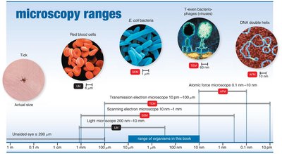
Units of Measurement in Microbiology
Micrometers and Nanometers
Micrometers (µm): 1 µm = 10-6 meters
Nanometers (nm): 1 nm = 10-9 meters
Microorganisms are typically measured in these units due to their small size.
Conversion: 1 µm = 1000 nm
Example: A typical bacterium is about 1–10 µm in length, while viruses range from 20–300 nm.
Microscopy: The Instruments
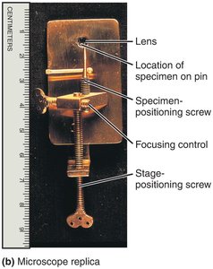
Simple and Compound Microscopes
Simple microscope: Contains a single lens, similar to a magnifying glass but with higher quality.
Compound microscope: Uses multiple lenses (objective and ocular) to achieve higher magnification and resolution.
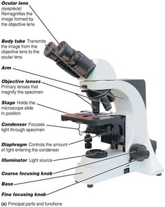
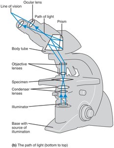
Path of Light in a Compound Microscope
Light passes from the illuminator through the condenser, specimen, objective lens, body tube, and ocular lens to the eye.
Total magnification: Product of the magnification of the objective and ocular lenses.
Resolution: The ability to distinguish two points as separate; higher resolution allows for finer detail.
Shorter wavelengths of light provide greater resolution.
Formula:
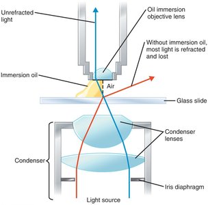
Refractive Index and Immersion Oil
Refractive index: Measure of how much a substance bends light.
Immersion oil is used with high-power objectives to reduce light refraction and increase resolution.
Types of Light Microscopy
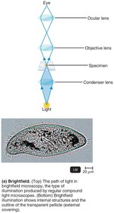
Brightfield Microscopy
Dark objects are visible against a bright background.
Most common type; best for stained specimens.
Unstained cells may be difficult to see due to low contrast.
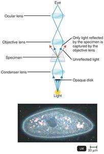
Darkfield Microscopy
Light objects are visible against a dark background.
Uses an opaque disk to block direct light; only reflected light enters the objective lens.
Useful for observing live, unstained microorganisms (e.g., Treponema pallidum).
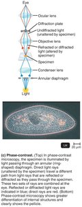
Phase-Contrast Microscopy
Enhances contrast of transparent specimens without staining.
Combines direct and diffracted light rays to visualize internal structures in living cells.
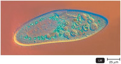
Differential Interference Contrast (DIC) Microscopy
Similar to phase-contrast but uses two beams and prisms for higher contrast and color.
Produces three-dimensional, brightly colored images.
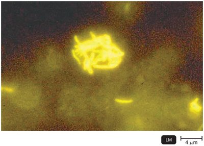
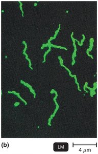
Fluorescence Microscopy
Uses UV light to excite fluorescent dyes (fluorochromes) that emit visible light.
Cells may be naturally fluorescent or stained with fluorochromes.
Immunofluorescence uses antibodies tagged with fluorochromes for specific detection of pathogens.
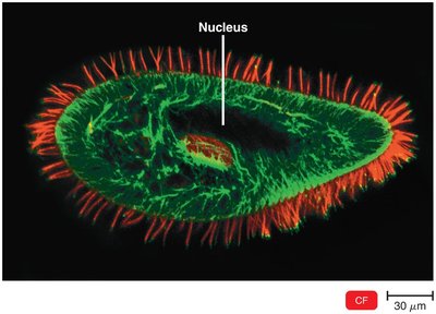
Confocal Microscopy
Uses fluorochromes and a laser to scan specimens in thin planes, producing clear two-dimensional images.
Computer reconstruction allows for three-dimensional imaging.
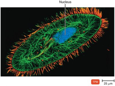
Two-Photon Microscopy
Uses two photons of long-wavelength light to excite fluorochromes.
Allows imaging of living cells up to 1 mm deep and tracking of cell activity in real time.
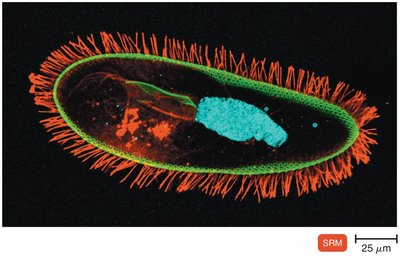
Super-Resolution Light Microscopy
Uses two laser beams to achieve resolution below the diffraction limit of light (as low as 1 nm).
Computer software reconstructs high-resolution images from scanned data.
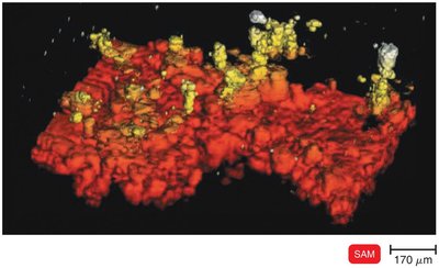
Scanning Acoustic Microscopy
Measures sound waves reflected from a specimen.
Used to study cells attached to surfaces, such as biofilms and cancer cells.
Resolution is about 1 µm.
Electron Microscopy
Principles and Types
Uses electron beams instead of light for much higher resolution (as small as 0.2 nm).
Essential for viewing viruses and internal cell structures.
Images are black and white but can be colorized digitally.
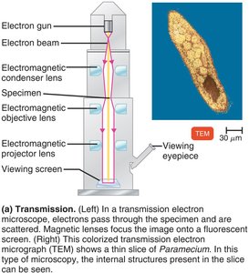
Transmission Electron Microscopy (TEM)
Electrons pass through ultrathin sections of a specimen.
Magnification: 10,000–10,000,000x; resolution: 0.2 nm.
Specimens must be fixed, dehydrated, and sectioned; preparation may introduce artifacts.
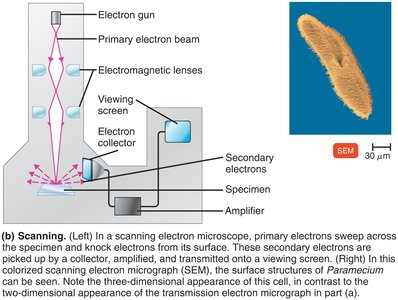
Scanning Electron Microscopy (SEM)
Electron beam scans the surface; secondary electrons are collected to form a 3D image.
Magnification: 1,000–500,000x; resolution: 0.5 nm.
Provides detailed surface views of specimens.
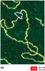
Scanned-Probe Microscopy
Scanning Tunneling Microscopy (STM)
Uses a tungsten probe to scan the surface at atomic resolution.
Can visualize molecules such as DNA without special preparation.
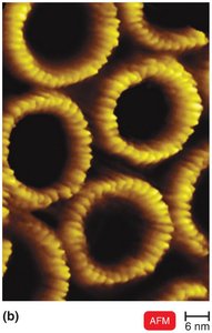
Atomic Force Microscopy (AFM)
Uses a metal-and-diamond probe to scan the specimen, producing 3D images at near-atomic detail.
Preparation of Specimens for Light Microscopy
Staining and Fixation
Staining: Coloring microorganisms with dyes to emphasize structures.
Smear: Thin film of specimen spread on a slide.
Fixation: Attaches and kills microorganisms, preserving structure (by heat or methanol).
Types of Dyes
Basic dyes: Chromophore is a cation (e.g., crystal violet, methylene blue); stains bacterial cells (negatively charged).
Acidic dyes: Chromophore is an anion (e.g., eosin, acid fuchsin); stains background (negative staining).
Simple Stains
Use a single basic dye to highlight the entire microorganism.
Examples: methylene blue, carbolfuchsin, crystal violet, safranin.
A mordant may be used to intensify the stain or enlarge structures.
Differential Stains
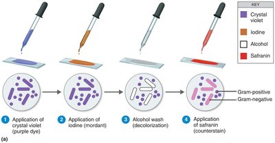
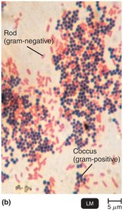
Gram Stain
Distinguishes between gram-positive (thick peptidoglycan, purple) and gram-negative (thin peptidoglycan, outer membrane, pink/red) bacteria.
Steps:
Step | Gram-Positive | Gram-Negative |
|---|---|---|
Primary Stain: Crystal Violet | Purple | Purple |
Mordant: Iodine | Purple | Purple |
Decolorizer: Alcohol/Acetone | Purple | Colorless |
Counterstain: Safranin | Purple | Pink/Red |
Acid-Fast Stain
Identifies bacteria with waxy cell walls (e.g., Mycobacterium, Nocardia).
Acid-fast cells retain red dye (carbolfuchsin) after acid-alcohol wash; non–acid-fast cells are blue after counterstaining with methylene blue.
Step | Acid-Fast | Non–Acid-Fast |
|---|---|---|
Primary Stain: Carbolfuchsin | Red | Red |
Decolorizer: Acid-Alcohol | Red | Colorless |
Counterstain: Methylene Blue | Red | Blue |
Special Stains
Capsule Stain
Capsules do not accept most dyes; negative staining with India ink or nigrosin highlights the capsule as a halo around the cell.
Endospore Stain
Endospores are resistant, dormant structures; stained with malachite green (with heat), counterstained with safranin.
Endospores appear green within red/pink cells.
Flagella Stain
Flagella are stained with a mordant and carbolfuchsin to visualize their number and arrangement.
Summary Table: Types of Microscopy and Their Uses
Microscopy Type | Principle | Best For |
|---|---|---|
Brightfield | Light passes through specimen | Stained cells, general morphology |
Darkfield | Only reflected light enters lens | Live, unstained cells |
Phase-Contrast | Combines direct and diffracted light | Internal structures of live cells |
DIC | Two beams, prisms for 3D effect | 3D, colored images of live cells |
Fluorescence | UV light excites fluorochromes | Specific detection, immunofluorescence |
Confocal | Laser scans thin planes | 3D reconstructions |
Electron (TEM/SEM) | Electron beams | Viruses, ultrastructure, surfaces |
Scanning Probe (STM/AFM) | Physical probe scans surface | Atomic/molecular detail |
Additional info: This guide covers the essential microscopy and staining techniques used in microbiology, providing foundational knowledge for laboratory practice and clinical diagnostics.
