 Back
BackChapter 3 - Observing Microorganisms Through a Microscope: Principles and Techniques
Study Guide - Smart Notes
 Tailored notes based on your materials, expanded with key definitions, examples, and context.
Observing Microorganisms Through a Microscope
Introduction to Microscopy in Microbiology
Microscopy is fundamental to microbiology, enabling the visualization and study of microorganisms that are otherwise invisible to the naked eye. This chapter explores the principles, types, and applications of various microscopes and staining techniques essential for observing microbial life.
Units of Measurement in Microbiology
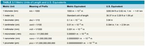
Metric Units and Their Relevance
Microorganisms are typically measured in micrometers (µm) and nanometers (nm).
Key conversions:
1 meter (m) = 100 centimeters (cm)
1 cm = 10 millimeters (mm)
1 mm = 1,000 micrometers (µm)
1 µm = 1,000 nanometers (nm)
1 nm = 0.001 µm
Metric Unit | Meaning of Prefix | Metric Equivalent | U.S. Equivalent |
|---|---|---|---|
Meter (m) | — | 1 m | 39.37 in. or 1.09 yd |
Centimeter (cm) | centi = 1/100 | 0.01 m | 0.39 in. |
Millimeter (mm) | milli = 1/1,000 | 0.001 m | 0.039 in. |
Micrometer (µm) | micro = 1/1,000,000 | 0.000001 m | 0.000039 in. |
Nanometer (nm) | nano = 1/1,000,000,000 | 0.000000001 m | 0.000000039 in. |
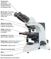
Principles of Microscopy
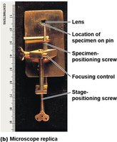
Simple and Compound Microscopes
Simple microscope: Contains a single lens, similar to a magnifying glass but with higher magnification.
Compound microscope: Uses multiple lenses (objective and ocular) to achieve higher magnification and resolution.
Total magnification:
Microscope Ranges and Magnification
Different microscopes are used to observe objects of varying sizes, from cells to viruses and molecules.
Resolution increases with decreasing wavelength of illumination.
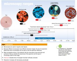
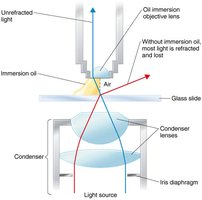
Resolution and Refractive Index
Resolution: The ability to distinguish two points as separate entities. Higher resolution allows for clearer images of small structures.
Refractive index: A measure of how much a substance bends light. Immersion oil is used to reduce light refraction and improve resolution at high magnification.
Types of Light Microscopy
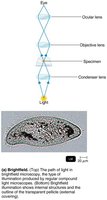
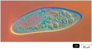
Brightfield Microscopy
Brightfield microscopy is the most common form, where dark objects are visible against a bright background. It is suitable for stained specimens but not for very small objects like viruses.
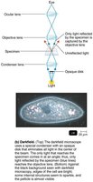
Darkfield Microscopy
Darkfield microscopy enhances the contrast in unstained specimens. Light objects are visible against a dark background, making it useful for detecting small or thin organisms.

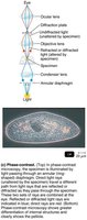
Phase-Contrast Microscopy
Phase-contrast microscopy allows for the detailed observation of living, unstained cells by amplifying differences in refractive index within the specimen. It is ideal for studying internal structures.
Differential Interference Contrast (DIC) Microscopy
DIC microscopy uses two light beams and prisms to produce high-contrast, colored, three-dimensional images of specimens, enhancing visualization of fine details.
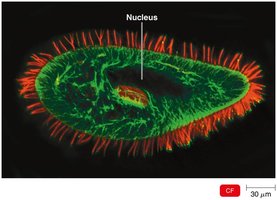
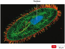
Fluorescence Microscopy
Fluorescence microscopy uses UV light to excite fluorescent dyes (fluorochromes) bound to specific cell components, causing them to emit visible light. It is widely used in diagnostics and research, especially for immunofluorescence techniques.
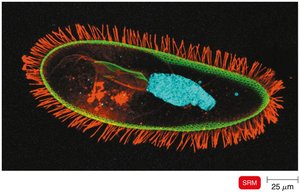
Confocal and Two-Photon Microscopy
Confocal microscopy: Uses lasers and fluorochromes to construct three-dimensional images by scanning successive planes of a specimen.
Two-photon microscopy: Uses two photons of long-wavelength light to excite dyes, allowing imaging of living cells up to 1 mm deep.
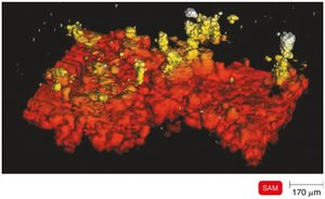
Super-Resolution and Scanning Acoustic Microscopy
Super-resolution microscopy: Uses two laser beams to achieve nanometer-scale resolution, allowing visualization of structures below the diffraction limit of light.
Scanning acoustic microscopy: Uses sound waves to visualize cells attached to surfaces, such as biofilms or cancer cells.
Electron Microscopy
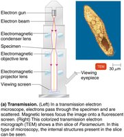
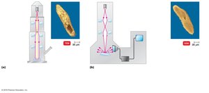
Transmission Electron Microscopy (TEM)
TEM passes electrons through ultrathin sections of a specimen, revealing internal structures at very high magnification and resolution. Specimens are often stained with heavy metals for contrast.
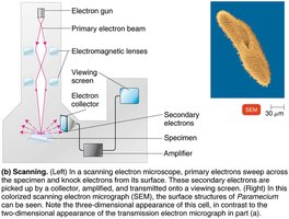
Scanning Electron Microscopy (SEM)
SEM scans the surface of a specimen with electrons, producing detailed three-dimensional images of surface structures. It is ideal for studying the morphology of cells and viruses.
Comparison of TEM and SEM
Microscope Type | Distinguishing Features | Typical Image | Principal Uses |
|---|---|---|---|
Transmission (TEM) | Electrons pass through ultrathin sections | Internal cell structures | Study internal ultrastructure |
Scanning (SEM) | Electrons scan the surface | Surface morphology | Study surface features |
Scanned-Probe Microscopy
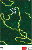
Scanning Tunneling Microscopy (STM)
STM uses a tungsten probe to scan the surface of a specimen, revealing atomic-level details. It is used for imaging DNA and other molecules.
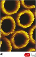
Atomic Force Microscopy (AFM)
AFM uses a metal-and-diamond probe to scan the specimen, producing three-dimensional images at near-atomic resolution. It is valuable for studying biological molecules and cell surfaces.
Preparation of Specimens for Light Microscopy
Staining and Smear Preparation
Staining: Coloring microorganisms with dyes to emphasize structures.
Smear: A thin film of microorganisms spread on a slide and fixed by heat or chemicals.
Live, unstained specimens are used to study cell behavior but have low contrast.
Types of Dyes
Basic dyes: Chromophore is a cation; stains bacterial cells (which are negatively charged).
Acidic dyes: Chromophore is an anion; stains the background (negative staining).
Simple Stains
Use a single basic dye to highlight the entire microorganism, making cell shapes and structures visible.
A mordant may be used to intensify the stain or enlarge structures.
Differential Stains
Used to distinguish between different groups of bacteria.
Common types: Gram stain and acid-fast stain.
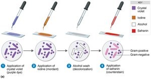
Gram Stain
The Gram stain differentiates bacteria based on cell wall structure:
Application of crystal violet (primary stain)
Application of iodine (mordant)
Alcohol wash (decolorization)
Application of safranin (counterstain)
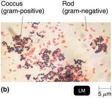
Gram-positive bacteria: Thick peptidoglycan wall, retain crystal violet, appear purple.
Gram-negative bacteria: Thin peptidoglycan wall and outer membrane, lose crystal violet, appear red/pink after safranin.
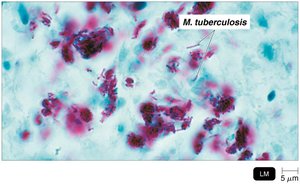
Acid-Fast Stain
Binds only to bacteria with waxy cell walls (e.g., Mycobacterium, Nocardia).
Acid-fast bacteria retain the primary stain even after acid-alcohol decolorization.
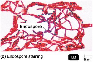
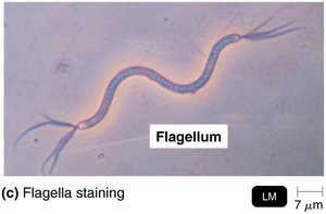
Special Stains
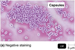
Capsule stain: Negative staining to visualize gelatinous capsules as halos around cells.
Endospore stain: Detects resistant, dormant structures within cells using malachite green and safranin.
Flagella stain: Uses a mordant and carbolfuchsin to thicken and visualize flagella.
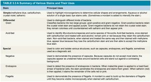
Summary Table: Stains and Their Uses
Stain | Principal Uses |
|---|---|
Simple | Highlight cell shapes and arrangements |
Differential (Gram, Acid-fast) | Distinguish between types of bacteria |
Special (Capsule, Endospore, Flagella) | Visualize specific structures |
Conclusion
Understanding the principles and applications of microscopy and staining is essential for the study of microorganisms. Mastery of these techniques allows microbiologists to identify, classify, and investigate the structure and function of diverse microbial life forms.
