 Back
BackObserving Microorganisms Through a Microscope: Principles and Techniques
Study Guide - Smart Notes
 Tailored notes based on your materials, expanded with key definitions, examples, and context.
Observing Microorganisms Through a Microscope
Units of Measurement
Microorganisms are extremely small and require specialized units for measurement. Understanding these units is essential for interpreting microscopic observations.
Micrometer (μm): 1 μm = 10−6 meters.
Nanometer (nm): 1 nm = 10−9 meters.
1 μm = 1,000 nm.
Microorganisms such as bacteria are typically measured in micrometers, while viruses are measured in nanometers.
Microscopy: The Instruments
Microscopy has revealed the structure and function of microorganisms. There are several types of microscopes, each with unique features and applications.
Simple Microscope: Contains a single lens.
Compound Microscope: Contains multiple lenses for higher magnification and resolution.
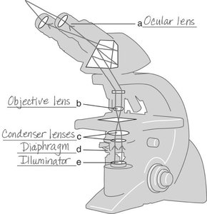
Path of Light Through a Compound Microscope
The compound light microscope is the most common instrument in microbiology. It uses visible light and a series of lenses to magnify specimens.
Light passes from the illuminator through the condenser lenses, then through the diaphragm, the specimen, the objective lens, and finally the ocular lens.
Total Magnification and Resolution
Magnification and resolution are critical concepts in microscopy.
Total Magnification: Calculated by multiplying the magnification of the objective lens by that of the ocular lens.
Resolution (Resolving Power): The ability to distinguish two points as separate. The maximum resolution of a compound light microscope is 0.2 μm.
Example: If the objective lens is 40× and the ocular lens is 10×, total magnification is 400×.
Types of Light Microscopy
Different types of light microscopy are used to observe various features of microorganisms.
Brightfield Microscopy: Used for stained specimens; provides a clear image of the specimen against a bright background.
Darkfield Microscopy: Useful for observing unstained, live specimens; shows a light silhouette against a dark background.
Phase-Contrast Microscopy: Allows detailed observation of living organisms by enhancing contrast without staining.
Differential Interference Contrast (DIC) Microscopy: Produces colored, three-dimensional images of living cells.
Fluorescence Microscopy: Uses fluorochromes and ultraviolet light to visualize specimens; important in diagnostic immunofluorescence techniques.
Confocal Microscopy: Uses fluorescent dyes and short-wavelength light to produce three-dimensional images.
Two-Photon Microscopy (TPM): Uses long-wavelength light to image live specimens stained with fluorescent dyes.
Super-Resolution Light Microscopy: Uses lasers and computer processing to achieve higher resolution images.
Scanning Acoustic Microscopy (SAM): Uses sound waves to study living cells attached to surfaces, such as biofilms.
Electron and Scanned-Probe Microscopy
Electron and scanned-probe microscopes provide much higher resolution than light microscopes.
Electron Microscopy: Uses electron beams instead of light and electromagnets instead of glass lenses.
Transmission Electron Microscope (TEM): Used for viewing thin sections of specimens; provides high magnification (10,000–10,000,000×) and resolution (10 pm).
Scanning Electron Microscope (SEM): Used for three-dimensional views of surfaces; magnification up to 500,000×, resolution 10 nm.
Scanning Tunneling Microscopy (STM) and Atomic Force Microscopy (AFM): Produce three-dimensional images of molecular surfaces using electric current or force detection.
Preparation of Specimens for Light Microscopy
Proper specimen preparation is essential for effective microscopic examination.
Staining: Coloring microorganisms with dyes to increase visibility of structures.
Fixing: Using heat or alcohol to kill and adhere microorganisms to the slide.
Smear: A thin film of material spread on a slide for examination.
Types of Stains
Basic Dyes: Positively charged; stain bacterial cells (which are negatively charged).
Acidic Dyes: Negatively charged; stain the background, producing a negative stain.
Simple Stain: Uses a single basic dye to highlight the entire microorganism.
Mordant: A substance that enhances the binding of the stain to the specimen.
Differential Stains
Gram Stain: Differentiates bacteria into gram-positive (purple) and gram-negative (pink) based on cell wall structure.
Acid-Fast Stain: Identifies acid-fast bacteria (e.g., Mycobacterium); acid-fast cells retain red dye, non-acid-fast cells appear blue.
Special Stains
Capsule Stain: Visualizes microbial capsules using negative staining.
Endospore Stain: Identifies bacterial endospores, which are resistant to standard staining.
Flagella Stain: Visualizes bacterial flagella for motility studies.
Applications and Interpretation of Microscopy
Microscopy is essential for diagnosing infections, studying microbial structure, and understanding microbial diversity.
Clinical Application: Different stains are used for different diagnostic purposes (e.g., Gram stain for pneumonia, acid-fast stain for tuberculosis).
Interpretation: Proper technique and understanding of staining reactions are crucial for accurate diagnosis.
Summary Table: Comparison of Microscopy Techniques
Microscopy Type | Key Feature | Application |
|---|---|---|
Brightfield | Stained cells, bright background | General observation |
Darkfield | Light silhouette, dark background | Unstained, live cells |
Phase-Contrast | Enhanced contrast, live cells | Intracellular structures |
DIC | 3D, colored images | Living cells |
Fluorescence | Fluorescent dyes, UV light | Immunofluorescence |
Confocal | 3D images, short-wavelength light | Cell imaging |
TEM | Thin sections, high magnification | Internal structures |
SEM | 3D surface images | Surface morphology |
Key Terms and Definitions
Magnification: The process of enlarging the appearance of an object.
Resolution: The ability to distinguish two points as separate entities.
Mordant: A chemical that helps fix a dye to a specimen.
Counterstain: A secondary stain used to provide contrast in differential staining.
Acid-Fast: Refers to bacteria that retain certain dyes after being washed with acid-alcohol.
Example Applications
Gram Stain: Used to guide antibiotic therapy by distinguishing between gram-positive and gram-negative bacteria.
Acid-Fast Stain: Essential for diagnosing tuberculosis caused by Mycobacterium species.
Capsule Stain: Identifies bacteria with protective capsules, important for understanding pathogenicity.
Additional Info:
Immersion oil is used with high-power objectives to reduce light refraction and improve resolution.
Heat-fixing is not used for capsule stains to avoid destroying the capsule structure.
Proper interpretation of staining results is critical for accurate microbial identification and diagnosis.
