 Back
Observing Microorganisms Through a Microscope: Principles, Techniques, and Applications
Study Guide - Smart Notes
 Tailored notes based on your materials, expanded with key definitions, examples, and context.
Observing Microorganisms Through a Microscope
Introduction
Microscopy is fundamental to microbiology, allowing scientists to visualize organisms too small to be seen with the naked eye. This chapter explores the principles of microscopy, the types of microscopes used, and the staining techniques essential for observing microbial structure and function.
Units of Measurement in Microbiology
Metric Units and Conversions
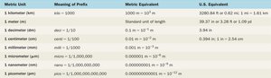
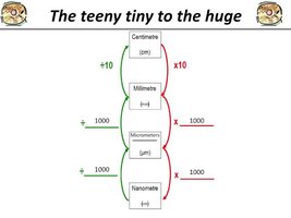
Microorganisms are measured using the metric system, primarily in micrometers (µm) and nanometers (nm). Understanding these units and their conversions is essential for interpreting microscopic observations.
Micrometer (µm): 1 µm = 10-6 meters
Nanometer (nm): 1 nm = 10-9 meters
1 µm = 1,000 nm
1 nm = 0.001 µm
1 cm = 0.01 m
Microbial Size Range
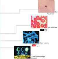
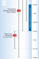
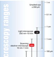
Bacteria typically range from 2–8 µm, with some as small as 200 nm. Viruses are much smaller, ranging from about 20 nm to 250 nm. The size of the specimen determines the appropriate microscope for observation.
Principles of Microscopy
Key Concepts
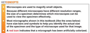
Microscopes magnify small objects, allowing for detailed observation of microorganisms.
The type of microscope used depends on the size and detail required for the specimen.
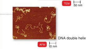
Micrographs often include scale bars and microscope type indicators for reference.
Magnification and Resolution
Magnification is the process of enlarging the appearance of an object. In compound light microscopes, total magnification is calculated by multiplying the magnification of the ocular lens by that of the objective lens:
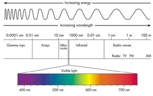
Resolution (resolving power) is the ability to distinguish two points as separate entities. The maximum resolution of a light microscope is about 0.2 µm, limited by the wavelength of visible light.
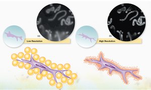
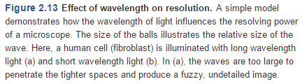
Key Point: Shorter wavelengths of light provide better resolution.
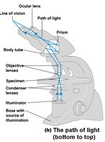
Path of Light in a Compound Microscope
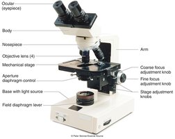
Light passes through a series of lenses to magnify the specimen. The main components include the light source, condenser, objective lens, and ocular lens.
Refractive Index and Immersion Oil
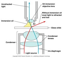
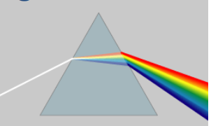
The refractive index measures how much a substance bends light. Using immersion oil (with a refractive index similar to glass) between the slide and the objective lens reduces light refraction, allowing more light to enter the lens and improving image clarity at high magnification.
Contrast
Contrast is the difference in light intensity between the specimen and the background. Staining increases contrast by changing the refractive index of the specimen, making structures more visible.
Types of Light Microscopes
Brightfield Microscopy
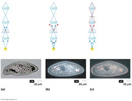
This is the most common type of light microscopy. It produces a dark image against a bright background. Staining is often used to enhance contrast.
Darkfield Microscopy
Darkfield microscopy makes light objects visible against a dark background, ideal for observing live, unstained specimens such as spirochetes.
Fluorescence Microscopy
Fluorescence microscopes use ultraviolet (UV) light to excite fluorescent dyes (fluorochromes) that emit visible light. This technique is especially useful for diagnostic applications, such as immunofluorescence, where antibodies labeled with fluorochromes bind to specific antigens.
Electron Microscopy
Principles and Types
Electron microscopes use beams of electrons instead of light, allowing for much higher resolution (down to the nanometer or even picometer scale). Images are always black and white but may be colorized for clarity.
Transmission Electron Microscope (TEM): Electrons pass through ultrathin sections of specimens, revealing internal structures in high detail (not 3D).
Scanning Electron Microscope (SEM): Electrons scan the surface of a specimen, producing detailed 3D images of surfaces.
Staining Techniques
Purpose of Staining
Staining enhances contrast in colorless cells, making cellular structures visible under the microscope. The basic steps are: smear, air-dry, and heat-fix.
Types of Dyes
Basic dyes: Positively charged chromophores; stain negatively charged cell components.
Acidic dyes: Negatively charged chromophores; used for negative staining (background staining).
Simple Stains
Simple stains use a single basic dye to reveal cell shape, size, and arrangement. Sometimes a mordant is used to enhance binding of the dye to the cell wall.
Differential Stains
Differential stains distinguish between different types of bacteria or structures. The two most important are the Gram stain and the acid-fast stain.
Gram Stain
The Gram stain differentiates bacteria based on cell wall structure:
Application of crystal violet (primary stain)
Application of iodine (mordant)
Alcohol wash (decolorization)
Application of safranin (counterstain)
Gram-positive cells: Remain purple after staining. Gram-negative cells: Appear pink/red after counterstaining.
Acid-Fast Stain
Used to identify bacteria with waxy cell walls (e.g., Mycobacterium and Nocardia). Acid-fast cells retain the primary stain (carbol fuchsin) after acid-alcohol treatment and appear reddish-purple; non–acid-fast cells are counterstained blue.
Special Stains
Endospore stain: Uses heat to drive malachite green into endospores; counterstain with safranin. Endospores appear green, vegetative cells red.
Capsule (negative) stain: Stains the background with acidic dye and the cell with a basic dye; capsules appear as clear halos.
Flagella stain: Uses a mordant to thicken flagella for visibility.
Summary Table: Types of Microscopes and Their Uses
Microscope Type | Principle | Resolution | Main Uses |
|---|---|---|---|
Brightfield | Light passes through specimen; staining enhances contrast | ~0.2 µm | General observation of stained cells |
Darkfield | Light reflected off specimen; dark background | ~0.2 µm | Live, unstained specimens (e.g., spirochetes) |
Fluorescence | UV light excites fluorochromes | ~0.2 µm | Diagnostic immunofluorescence |
TEM | Electrons pass through ultrathin sections | ~2 nm | Internal cell structures, viruses |
SEM | Electrons scan specimen surface | ~10 nm | 3D surface images |
Practice and Application
Convert units as needed for interpreting micrographs (e.g., 10 µm = 10,000 nm).
Choose the appropriate microscope based on specimen size and required detail.
Apply the correct staining technique for the organism or structure of interest.
Additional info: Mastery of microscopy and staining is foundational for all laboratory work in microbiology, enabling the identification and study of bacteria, viruses, and other microorganisms.
