 Back
BackObserving Microorganisms Through a Microscope: Study Notes
Study Guide - Smart Notes
 Tailored notes based on your materials, expanded with key definitions, examples, and context.
Observing Microorganisms Through a Microscope
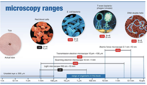
Units of Measurement
Microorganisms are measured using units much smaller than those visible to the unaided eye. Understanding these units is essential for interpreting microscopic observations.
Micrometers (μm): Used for bacteria; 1 μm = 10-6 meters.
Nanometers (nm): Used for viruses; 1 nm = 10-9 meters.
Unaided eye: Can see objects ≥ 200 μm; anything smaller requires magnification.
Example: Helicobacter pylori (H. pylori) is a bacterium causing stomach ulcers, typically measured in micrometers.
Microscopy: The Instruments
Microscopes are essential tools for observing microorganisms. They vary in complexity and magnification capabilities.
Simple microscope: Contains a single lens; pioneered by Anton van Leeuwenhoek.
Compound microscope: Uses multiple lenses for higher magnification.
Light Microscopy
Light microscopy utilizes visible light to observe specimens. Several types exist, each suited for different applications.
Compound light microscopy: Most common; uses objective and ocular lenses.
Darkfield microscopy: Enhances contrast for live, unstained specimens.
Phase-contrast microscopy: Allows detailed examination of living cells without staining.
Compound Light Microscopy
The compound microscope magnifies the image from the objective lens further with the ocular lens. Total magnification is calculated as:
Total Magnification:
Examples: 4x × 10x = 40x; 10x × 10x = 100x; 40x × 10x = 400x; 100x × 10x = 1000x (oil immersion).
Resolution (resolving power): The ability to distinguish two points as separate. Shorter wavelengths yield greater resolution. The limit for compound light microscopes is 0.2 μm.
Refractive Index: Measures light-bending ability. Immersion oil is used to reduce refraction and improve resolution.
Illumination Techniques
Brightfield: Dark objects visible against a bright background; unstained cells may lack contrast.
Darkfield: Light objects visible against a dark background; useful for live, unstained microorganisms such as Treponema pallidum (causes syphilis).
Phase-Contrast: Combines direct and diffracted light rays for detailed examination of living cells; no staining required.
Electron Microscopy
Electron microscopes use electron beams instead of light, providing much higher resolution for observing viruses and internal cell structures.
Transmission Electron Microscopy (TEM): Electrons pass through ultrathin specimen sections; magnification 10,000–10,000,000x.
Scanning Electron Microscopy (SEM): Electrons scan the specimen surface, producing 3D images; magnification 1,000–500,000x.
Preparing Smears for Staining
Staining is used to enhance contrast and visualize specific structures in microorganisms. Smears are thin films of material containing microorganisms spread on a slide.
Fixing: Attaches and kills microorganisms, preserving their structure.
Stains: Composed of positive and negative ions; the colored ion is called the chromophore.
Basic dye: Chromophore is a cation (e.g., crystal violet, methylene blue, safranin).
Acidic dye: Chromophore is an anion (e.g., nigrosine).
Negative staining: Stains the background, not the cell; uses acidic dyes.
Simple Stains
Simple stains use a single basic dye to highlight the entire microorganism, making cell shapes and structures visible.
Examples: Methylene blue, carbolfuchsin, crystal violet, safranin.
Mordant: Intensifies the stain.
Differential Stains
Differential stains distinguish between different types of bacteria. The most common are the Gram stain and Acid-fast stain.
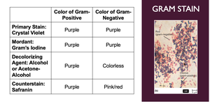
Gram Stain
The Gram stain classifies bacteria as gram-positive or gram-negative based on cell wall structure. It is a critical technique in medical microbiology for identifying bacteria and guiding treatment.
Gram-positive: Thick peptidoglycan cell wall; stains purple.
Gram-negative: Thin peptidoglycan cell wall and outer membrane; stains pink/red.
Gram stains: Most consistent on young, actively growing bacteria.

Step | Color of Gram-Positive | Color of Gram-Negative |
|---|---|---|
Primary Stain: Crystal Violet | Purple | Purple |
Mordant: Gram's Iodine | Purple | Purple |
Decolorizing Agent: Alcohol or Acetone-Alcohol | Purple | Colorless |
Counterstain: Safranin | Purple | Pink/red |
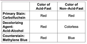
Acid-Fast Stain
The Acid-fast stain is used to identify bacteria with waxy cell walls, such as Mycobacterium and Nocardia. These bacteria retain the primary stain even after decolorization with acid-alcohol.
Acid-fast bacteria: Stain red.
Non-acid-fast bacteria: Stain blue after counterstaining.
Steps: Carbolfuchsin → acid-alcohol → methylene blue.
Step | Color of Acid-Fast | Color of Non–Acid-Fast |
|---|---|---|
Primary Stain: Carbolfuchsin | Red | Red |
Decolorizing Agent: Acid-Alcohol | Red | Colorless |
Counterstain: Methylene Blue | Red | Blue |
Special Stains
Special stains are used to highlight specific structures within microorganisms, such as capsules and endospores.
Negative Staining for Capsules
Capsules: Gelatinous coverings that do not accept most dyes.
Negative staining: Uses India ink or nigrosine to stain the background, making the capsule appear as a halo.
Endospore Staining
Endospores: Resistant, dormant structures inside some cells; not stained by ordinary methods.
Schaeffer-Fulton method: Uses malachite green (with heat), water to decolorize, and safranin as a counterstain. Spores appear green within red or pink cells.
Summary Table: Staining Techniques
Below is a summary of the main staining techniques and their purposes:
Stain | Purpose | Key Features |
|---|---|---|
Simple Stain | Visualize cell shape and structure | Single basic dye |
Gram Stain | Distinguish Gram-positive and Gram-negative bacteria | Four-step process; color differences |
Acid-Fast Stain | Identify acid-fast bacteria | Retains red color after acid-alcohol |
Capsule Stain | Visualize capsules | Negative staining; halo effect |
Endospore Stain | Visualize endospores | Malachite green and safranin |
Additional info: Academic context was added to clarify the purpose and application of each staining technique, as well as to provide definitions and examples for key terms.
