 Back
Back(Chapter 3) Observing Microorganisms Through a Microscope: Study Notes for Microbiology Students
Study Guide - Smart Notes
 Tailored notes based on your materials, expanded with key definitions, examples, and context.
Observing Microorganisms Through a Microscope
Units of Measurement
Microorganisms are measured using units much smaller than those used for everyday objects. Understanding these units is essential for interpreting microscopic observations.
Micrometer (μm): 1 μm = 10-6 meters
Nanometer (nm): 1 nm = 10-9 meters
Conversion: 1 μm = 1000 nm
Microorganisms such as bacteria are typically measured in μm, while viruses are measured in nm.
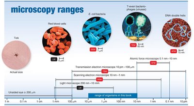
Example: E. coli bacteria are about 2 μm long, while a DNA double helix is about 2 nm in diameter.
Microscopy: The Instruments
Microscopes are essential tools in microbiology, allowing scientists to observe organisms too small to be seen with the naked eye. The earliest microscopes were simple, consisting of a single lens.
Simple microscope: Contains only one lens, similar to a magnifying glass but with higher magnification.
Anton van Leeuwenhoek: Developed early simple microscopes, enabling the first observations of microorganisms.
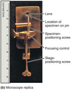
Example: Leeuwenhoek's microscope could magnify specimens up to 300x.
Light Microscopy
Light microscopes use visible light to illuminate specimens. Several types of light microscopy are used in microbiology, each with unique advantages.
Compound light microscopy: Uses multiple lenses to magnify specimens.
Darkfield microscopy: Enhances contrast for unstained, live specimens.
Phase-contrast microscopy: Reveals internal structures in living cells without staining.
Compound Light Microscopy
Compound microscopes use two sets of lenses: objective and ocular. The total magnification is the product of the magnifications of these lenses.
Total magnification:
Common magnifications: 100x (low power), 400x (high power), 1000x (oil immersion)
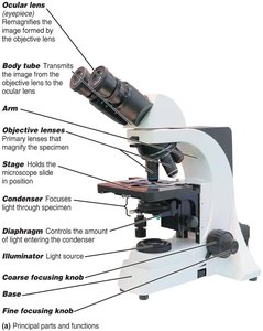
Example: Using a 40x objective and a 10x ocular lens gives a total magnification of 400x.
Resolution and Refractive Index
Resolution is the ability of a microscope to distinguish two points as separate entities. Higher resolution allows for clearer, more detailed images.
Resolving power: The minimum distance at which two points can be distinguished.
Shorter wavelengths of light provide greater resolution.
Limit of resolution for compound light microscopes: 0.2 μm
Refractive index: A measure of how much a medium bends light. Immersion oil is used to reduce refraction and increase resolution at high magnifications.
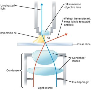
Example: Without immersion oil, light is refracted and lost, reducing image clarity.
Brightfield Illumination
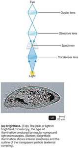
Brightfield microscopy is the standard method for observing stained specimens. It produces a bright background with dark objects.
Best for stained, fixed specimens.
Unstained cells may lack contrast and be difficult to see.
Darkfield Microscopy
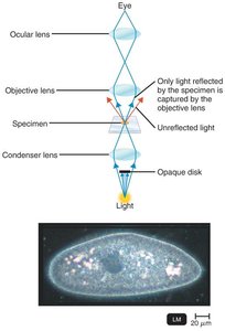
Darkfield microscopy enhances contrast by making specimens appear bright against a dark background. It is especially useful for observing live, unstained microorganisms.
Uses an opaque disk to block direct light.
Only light reflected by the specimen enters the objective lens.
Useful for viewing slender bacteria such as Treponema pallidum.
Phase-Contrast Microscopy
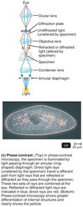
Phase-contrast microscopy allows for detailed examination of living cells and their internal structures without staining. It uses differences in refractive index to enhance contrast.
Combines direct and diffracted light rays to form an image.
Ideal for observing motility and internal structures.
Electron Microscopy
Electron microscopes use electron beams instead of light, providing much higher resolution. They are essential for studying viruses and internal cell structures.
Transmission Electron Microscopy (TEM): Electrons pass through thin sections of specimens, revealing internal structures.
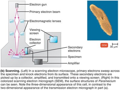
Scanning Electron Microscopy (SEM): Electrons scan the surface, producing three-dimensional images.
Transmission Electron Microscopy (TEM)
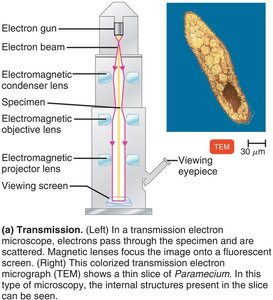
TEM provides detailed images of internal cell structures by passing electrons through ultrathin sections of specimens.
Magnification: 10,000–10,000,000x
Specimens are stained with heavy metals for contrast.
Preparation involves fixation, dehydration, and slicing.
Scanning Electron Microscopy (SEM)
SEM produces three-dimensional images by scanning the surface of specimens with electrons.
Magnification: 1,000–500,000x
Secondary electrons emitted from the specimen are collected and amplified.
Ideal for visualizing surface structures.
Preparing Smears for Staining
Staining is a critical technique in microbiology, used to enhance contrast and highlight specific structures in microorganisms.
Smear: A thin film of microorganisms spread on a slide.
Fixation: Attaches and kills microorganisms, preserving their structure.
Stains: Dyes with colored ions (chromophores); basic dyes have cationic chromophores, acidic dyes have anionic chromophores.
Bacterial cells are negatively charged, so basic dyes adhere to them.
Negative staining: Stains the background, not the cell, using acidic dyes.
Simple Stains
Simple stains use a single basic dye to highlight the entire microorganism, making cell shapes and structures visible.
Common dyes: methylene blue, carbolfuchsin, crystal violet, safranin
A mordant may be used to intensify the stain.
Differential Stains
Differential stains distinguish between different types of bacteria. The most important are the Gram stain and acid-fast stain.
Gram stain: Differentiates bacteria based on cell wall structure.
Acid-fast stain: Identifies bacteria with waxy cell walls.
Gram Stain
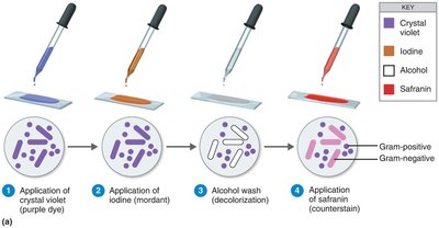
The Gram stain is a fundamental technique in medical microbiology, classifying bacteria as gram-positive or gram-negative based on cell wall composition.
Gram-positive: Thick peptidoglycan cell wall; stains purple.
Gram-negative: Thin peptidoglycan cell wall and outer membrane; stains pink/red.
Steps: Application of crystal violet (primary stain), iodine (mordant), alcohol (decolorizer), safranin (counterstain).
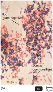
Example: Gram stains are used to identify bacteria in clinical specimens and guide treatment decisions.
Acid-Fast Stain
The acid-fast stain is used to identify bacteria with waxy cell walls, such as Mycobacterium and Nocardia. These bacteria retain the primary stain even after decolorization with acid-alcohol.
Acid-fast bacteria: Stain red with carbolfuchsin.
Non–acid-fast bacteria: Stain blue with methylene blue after decolorization.
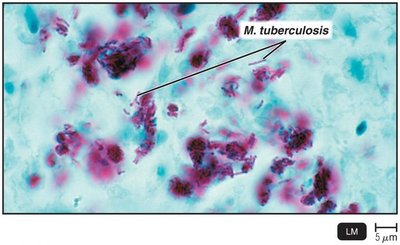
Example: Acid-fast staining is critical for diagnosing tuberculosis.
Special Stains
Special stains are used to highlight specific structures within microorganisms, such as capsules and endospores.
Capsule stain: Negative staining highlights the gelatinous capsule as a halo around the cell.
Endospore stain: Schaeffer-Fulton method stains endospores green and the rest of the cell red/pink.
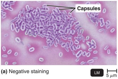
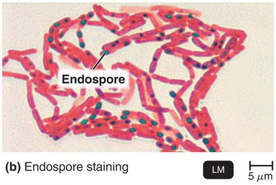
Example: Capsule staining is used to identify pathogenic bacteria with protective capsules; endospore staining is used to detect dormant, resistant structures in bacteria such as Bacillus and Clostridium.
