 Back
BackObserving Microorganisms Through a Microscope: Study Guide and Metric Conversions
Study Guide - Smart Notes
 Tailored notes based on your materials, expanded with key definitions, examples, and context.
Observing Microorganisms Through a Microscope
Units of Measurement in Microbiology
Microbiologists use specific units to measure microorganisms due to their small size. Understanding these units is essential for accurate observation and reporting.
Micrometer (µm): Commonly used for bacteria. 1 µm = 0.000001 m = 10-6 m
Nanometer (nm): Used for viruses and smaller structures. 1 nm = 0.000000001 m = 10-9 m
Examples:
Bacteria: 1–10 µm
Viruses: 20–300 nm
Remember: Bacteria are measured in micrometers, viruses in nanometers.
Metric System and Conversions
The metric system is used universally in microbiology for measuring microorganisms and their structures. Converting between units is a fundamental skill.
To convert nanometers to micrometers: divide by 1,000.
To convert micrometers to nanometers: multiply by 1,000.
To convert micrometers to millimeters: divide by 1,000.
To convert millimeters to micrometers: multiply by 1,000.
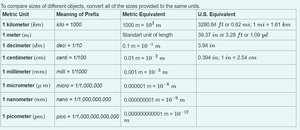
Example: 2,000 nm = 2 µm; 5 µm = 0.005 mm
Compound Light Microscope: Structure and Function
The compound light microscope is a fundamental tool in microbiology, allowing visualization of microorganisms using visible light and multiple lenses.
Illuminator: Light source
Condenser: Directs light through the specimen
Objective Lens: Primary magnification (4×, 10×, 40×, 100×)
Ocular Lens (Eyepiece): Remagnifies the image, usually 10×
Stage: Holds the slide
Coarse/Fine Adjustment Knobs: Focus the image
Oil Immersion Lens (100×): Used with oil to improve resolution
Total Magnification
Total magnification is calculated by multiplying the magnification of the objective lens by that of the ocular lens.
Formula:
Examples:
4× objective × 10× ocular = 40×
10× objective × 10× ocular = 100×
40× objective × 10× ocular = 400×
100× objective × 10× ocular = 1000×
Resolution
Resolution is the ability to distinguish two close objects as separate entities. It is a critical property for clarity in microscopy.
Better resolution = more detail
Shorter wavelength of light = better resolution
Light microscope resolution ≈ 0.2 µm
Key Point: Magnification makes things bigger; resolution makes things clearer.
Types of Light Microscopy
Different types of light microscopy are used to visualize microorganisms based on their properties and the information needed.
Brightfield: Standard; stained specimens
Darkfield: Live, unstained specimens; bright object on dark background (e.g., Treponema pallidum)
Phase-Contrast: Living, transparent cells; internal structures visible
DIC (Differential Interference Contrast): Nearly 3D images
Fluorescence: Uses fluorochromes; organisms glow
Confocal: Laser scanning; 3D images
Two-Photon: Live tissues; up to 1 mm deep
Electron Microscopy: TEM vs. SEM
Electron microscopes use electron beams for much higher resolution than light microscopes. There are two main types:
TEM (Transmission Electron Microscope): Internal structures; 2D image; electron beam passes through specimen
SEM (Scanning Electron Microscope): Surface structures; 3D image; electron beam scans specimen surface
Memory Trick: TEM = Through specimen (internal); SEM = Surface (external)
Staining Techniques in Microbiology
Staining increases contrast, making bacteria visible and allowing observation of shape, size, arrangement, and structures.
Basic Stains: Positively charged dyes (e.g., crystal violet, methylene blue); stain cells
Acidic Stains: Negatively charged dyes (e.g., eosin, nigrosin); stain background (negative stain)
Simple Stain: One dye; shows shape, size, arrangement. Differential Stain: Multiple dyes; distinguishes types of bacteria (e.g., Gram stain, acid-fast stain).
Gram Stain and Acid-Fast Stain
These are the most important differential stains in microbiology.
Gram Stain:
Crystal Violet
Iodine
Alcohol
Safranin
Results: Gram-positive = purple; Gram-negative = pink
Acid-Fast Stain:
Carbolfuchsin
Heat
Acid Alcohol
Methylene Blue
Results: Acid-fast = red/pink; Non-acid-fast = blue
Examples of Acid-Fast and Endospore-Producing Bacteria
Acid-Fast Bacteria: Mycobacterium tuberculosis (tuberculosis), Mycobacterium leprae (leprosy), Nocardia
Endospore Producers: Bacillus anthracis (anthrax), Clostridium tetani (tetanus), Clostridium botulinum (botulism), Clostridium difficile (severe diarrhea)
Key Fact: Most medically important endospore producers are Gram-positive rods: Bacillus (aerobic) and Clostridium (anaerobic).
Summary Table: Microscope Parts and Functions
Part | Function |
|---|---|
Ocular Lens (Eyepiece) | Remagnifies image; usually 10× |
Objective Lens | Primary magnification (4×, 10×, 40×, 100×) |
Condenser | Focuses light through specimen |
Diaphragm | Controls light entering condenser |
Illuminator | Light source |
Stage | Holds slide |
Stage Clips/Mechanical Stage | Holds slide in place |
Coarse Adjustment Knob | Rough focusing |
Fine Adjustment Knob | Sharp focus |
Revolving Nosepiece | Holds objective lenses |
Body Tube/Head | Maintains lens distance |
Arm | Supports upper parts |
Base | Bottom support |
Oil Immersion Lens (100×) | Improves resolution with oil |
Must-Know Facts for Exams
Bacteria = µm; Viruses = nm
Total Magnification = Objective × Ocular
Resolution = Ability to distinguish two objects
Light microscope resolution ≈ 0.2 µm
TEM = Internal structures; SEM = Surface structures
Basic stain = Cells colored; Acidic stain = Background colored
Simple stain = One dye; Differential stain = Multiple dyes
Gram Positive = Purple; Gram Negative = Pink
Gram stain: Crystal Violet, Iodine, Alcohol, Safranin
Acid-fast stain: Carbolfuchsin, Heat, Acid Alcohol, Methylene Blue
Acid-fast bacteria: Mycobacterium tuberculosis, Mycobacterium leprae, Nocardia
Endospore producers: Bacillus, Clostridium
