 Back
BackObserving Microorganisms Through a Microscope: Tools, Techniques, and Applications
Study Guide - Smart Notes
 Tailored notes based on your materials, expanded with key definitions, examples, and context.
Observing Microorganisms Through a Microscope
Introduction to Microbial Visualization
Microorganisms are too small to be seen with the unaided eye, requiring specialized tools and techniques for observation. Microscopy is fundamental in microbiology for studying the structure, function, and identification of microbes.
Tools and Techniques to Visualize Microbes
Microscopy: The Foundation of Microbial Observation
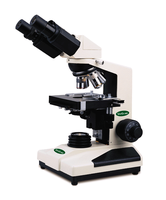
Microscope: An instrument that magnifies small objects, allowing detailed visualization of microorganisms.
Types of Microscopes: Simple (single lens) and compound (multiple lenses) microscopes.
Applications: Used to study bacteria, fungi, protozoa, and viruses (with electron microscopy).

A Sense of Scale
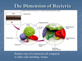
Understanding the size of microorganisms is crucial for selecting appropriate visualization techniques.
Unaided Vision: Can see objects > 200 μm (0.2 mm).
Common Bacteria: Typically 1–2 μm in size.
Viruses: Much smaller, often < 0.1 μm.
Types of Light Microscopes
Simple vs. Compound Microscopes
A simple microscope uses a single lens for magnification, while a compound microscope uses multiple lenses (objective and ocular) for higher magnification and resolution.
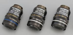
Compound Microscope: Most common in microbiology labs; allows for detailed observation of microbial cells.

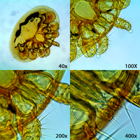
Magnification and Field of View
Magnification is the apparent enlargement of a specimen. As magnification increases, the field of view decreases, allowing for more detailed observation of smaller areas.
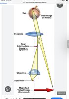
Principle of Refraction in Microscopy
Refraction is the change in direction of light as it passes through different materials, such as glass lenses. This principle is essential for focusing and magnifying images in microscopes.

Microscope Lenses and Image Formation
Microscope objectives use convex glass lenses to refract light and magnify specimens. The objective lens creates a real, magnified image, which is further enlarged by the ocular lens for viewing.
Resolution
Resolution is the ability to distinguish two points as separate entities. High resolution is essential for clear, detailed images. Magnification without adequate resolution results in blurry images.
Good Resolution: Distinct, separate points are visible.
Poor Resolution: Points appear merged or blurry.
Types of Light Microscopy
Brightfield (Compound Light) Microscopy
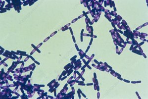
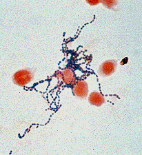
Brightfield microscopy uses visible light transmitted through the specimen. The specimen appears dark against a bright background. It is commonly used for stained specimens to observe cell size, shape, and arrangement.
Applications: Viewing stained bacteria, distinguishing features, and identifying live or dead cells.
Phase Contrast Microscopy
Phase contrast microscopy enhances contrast between light and dark areas by redirecting light using special filters. It is ideal for observing live, unstained cells and internal structures, especially in eukaryotes.
Applications: Viewing internal organelles and live cell dynamics without staining.
Fluorescence Microscopy
Fluorescence microscopy uses ultraviolet (UV) light to excite fluorescent molecules, which then emit visible light. This technique allows for the detection of specific targets within cells using fluorescent probes or antibodies.
Applications: Identifying specific proteins, pathogens, or nucleic acids in a mixture; disease diagnosis.
Example: Detecting rabies virus or specific bacteria in clinical samples.
Staining Techniques in Light Microscopy
Simple Stains
Simple stains use a single dye to color all cells the same, making it easier to observe cell size, shape, and arrangement.
Differential Stains
Differential stains use multiple dyes and a decolorizer step to distinguish between different types of cells or structures. Common examples include the Gram stain, endospore stain, and acid-fast stain.
Gram Stain: Differentiates bacteria based on cell wall structure (Gram-positive vs. Gram-negative).
Endospore Stain: Identifies bacterial endospores.
Acid-Fast Stain: Identifies Mycobacterium species (e.g., tuberculosis).
Electron Microscopy
Overview and Advantages
Electron microscopy uses a beam of electrons instead of light, allowing for much higher magnification and resolution. It is essential for studying viruses and fine cellular details.
Transmission Electron Microscopy (TEM): Electrons pass through ultra-thin specimens, revealing internal structures at very high resolution (up to 0.01 nm).
Scanning Electron Microscopy (SEM): Electrons scan the surface, producing detailed 3D images of specimen surfaces (resolution up to 10 nm).
Limitations: Cannot be used for live specimens; requires extensive preparation and specialized facilities.
Summary Table: Types of Microscopy
Microscope Type | Principle | Best Use | Resolution |
|---|---|---|---|
Brightfield (Compound Light) | Visible light through specimen | Stained cells, general observation | ~200 nm |
Phase Contrast | Enhances contrast via light phase shifts | Live, unstained cells | ~200 nm |
Fluorescence | UV light excites fluorescent dyes | Detecting specific targets | ~200 nm |
TEM | Electron beam through specimen | Internal structures, viruses | 0.01 nm |
SEM | Electron beam scans surface | Surface details, 3D images | 10 nm |
Key Terms and Concepts
Magnification: The process of enlarging the appearance of an object.
Resolution: The ability to distinguish two points as separate.
Refraction: Bending of light as it passes through different media.
Staining: Application of dyes to enhance contrast in microscopic images.
Electron Microscopy: Uses electrons for imaging, allowing for much higher resolution than light microscopy.
Sample Exam Questions
What is the main advantage of phase contrast microscopy over brightfield microscopy?
Why is fluorescence microscopy particularly useful in disease diagnosis?
Describe the main differences between TEM and SEM.
Explain why electron microscopes cannot be used to view live specimens.
