 Back
BackPathogenic Gram-Positive Bacteria: Structure, Diseases, and Clinical Management
Study Guide - Smart Notes
 Tailored notes based on your materials, expanded with key definitions, examples, and context.
Pathogenic Gram-Positive Bacteria
Structure and Physiology
Gram-positive pathogenic bacteria exhibit diverse morphologies and physiological traits. Two important genera discussed here are Corynebacterium and Mycobacterium. These bacteria are notable for their pleomorphism, non-endospore-forming nature, and unique cell wall components.
Pleomorphic, non-endospore-forming bacteria: These bacteria can vary in shape and do not produce endospores.
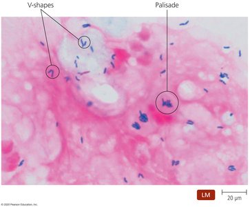
Snapping division: A distinctive method of cell division resulting in characteristic arrangements such as V-shapes and palisades.
Habitat: Ubiquitous on plants, animals, and humans, colonizing skin and mucosal surfaces.
Corynebacterium diphtheriae
Pathogenesis, Epidemiology, and Disease
Corynebacterium diphtheriae is the causative agent of diphtheria, a potentially fatal respiratory disease. The pathogenicity is primarily due to the production of diphtheria toxin.
Transmission: Person-to-person via respiratory droplets or skin contact.
Diphtheria toxin: Inhibits polypeptide synthesis in eukaryotic cells, leading to cell death.
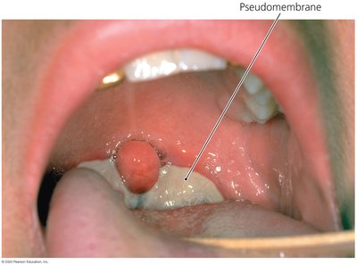
Pseudomembrane formation: A hallmark of diphtheria, which can cause airway obstruction and suffocation.
Non-toxigenic strains: Do not cause disease.
Diagnosis, Treatment, and Prevention
Diagnosis: Based on clinical presentation (pseudomembrane), confirmed by culture on potassium-tellurite agar, Loffler’s medium, or Elek test.
Treatment: Antitoxin administration, antibiotics (penicillin or erythromycin), and surgical intervention if airway is blocked.
Prevention: Immunization (DTaP vaccine) is the most effective preventive measure.
Mycobacterium: Structure and Physiology
Unique Cell Wall and Acid-Fastness
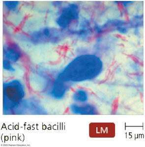
Members of the genus Mycobacterium are non-endospore-forming, acid-fast bacteria with a cell wall rich in mycolic acid. This confers several unique properties:
Slow growth due to the complex cell wall.
Resistance to desiccation, Gram-staining, detergents, and many antimicrobial drugs.
Intracellular survival and protection from lysis after phagocytosis.
Mycobacterium tuberculosis and Tuberculosis (TB)
Pathogenesis and Disease Forms
Mycobacterium tuberculosis causes tuberculosis, a chronic respiratory disease. The bacterium is not highly virulent but can persist in the host for years.
Primary TB: Initial infection, may be asymptomatic or symptomatic.
Secondary (reactivated) TB: Reactivation of dormant bacteria, often due to immunosuppression.
Disseminated TB: Spread of infection to multiple organs, causing varied symptoms.
Pathogenicity: Mycobacteria prevent fusion of lysosomes with phagosomes in macrophages, allowing intracellular survival.
Diagnosis, Treatment, and Prevention
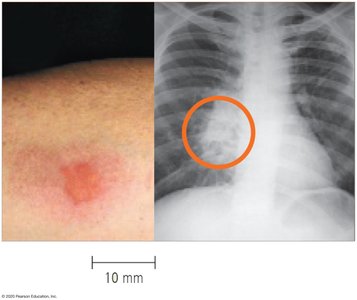
Diagnosis: Tuberculin skin test (Mantoux test) for exposure, chest X-rays for active disease.
Treatment: Combination therapy with isoniazid (INH), rifampin, ethambutol or streptomycin, and pyrazinamide for several months.
Prevention: BCG vaccine in endemic areas, avoiding exposure to respiratory droplets.
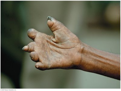
Mycobacterium leprae and Leprosy (Hansen’s Disease)
Pathogenesis, Epidemiology, and Disease Forms
Mycobacterium leprae causes leprosy, which affects cooler regions of the body. The bacterium cannot be cultured in cell-free media; armadillos and humans are the only known hosts.
Tuberculoid leprosy: Nonprogressive, due to strong cell-mediated immunity.
Lepromatous leprosy: More severe, due to weak cell-mediated immunity.
Transmission: Person-to-person contact or breaks in the skin.
Diagnosis, Treatment, and Prevention
Diagnosis: Based on clinical signs and symptoms.
Treatment: Combination of antimicrobial drugs; sometimes lifelong therapy is required.
Prevention: Limiting exposure and BCG vaccination provide some protection.
Other Mycobacterial Infections
Mycobacterium avium-intracellulare Complex
This complex is the most common mycobacterial infection among AIDS patients in the United States. Infection occurs via ingestion of contaminated food or water and can affect nearly every organ, often resulting in massive organ failure. Treatment is challenging due to the disseminated nature of the infection.
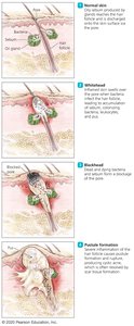
Cutibacterium acnes
Structure, Pathogenesis, and Disease
Cutibacterium acnes (formerly Propionibacterium acnes) is a small, anaerobic Gram-positive rod commonly found on human skin. It is the primary cause of acne in adolescents and young adults.
Pathogenesis: Utilizes sebum as a nutrient source, leading to inflammation and acne formation.
Opportunistic infections: May occur in other body sites, especially in immunocompromised individuals.
Treatment: Most cases are self-limiting; antimicrobials may be used for severe cases.
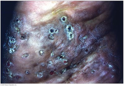
Nocardia and Actinomyces: Fungal-Like Bacteria
Nocardia asteroides
Nocardia asteroides is a soil-dwelling, opportunistic pathogen that can cause pulmonary, cutaneous, and central nervous system infections, especially in immunocompromised patients.
Transmission: Inhalation or introduction into wounds.
Mycetoma formation: Chronic, localized infection with draining sinuses.
Treatment: Prolonged course of sulfonamides; prognosis is poor in immunocompromised hosts.
Prevention: Avoid exposure to contaminated soil.
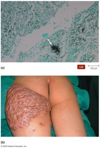
Actinomyces
Actinomyces species are normal microbiota of human mucous membranes but can cause opportunistic infections when mucosal barriers are breached.
Pathogenesis: Entry through breaks in mucous membranes leads to abscesses connected by sinus tracts.
Sites of infection: Respiratory, gastrointestinal, urinary, and female genital tracts.
Treatment: Surgical removal of infected tissue and prolonged penicillin therapy.
Prevention: Good oral hygiene and prophylactic antimicrobials if mucosal injury occurs.
Summary Table: Key Pathogenic Gram-Positive Bacteria
Bacterium | Main Disease(s) | Key Features | Diagnosis | Treatment | Prevention |
|---|---|---|---|---|---|
Corynebacterium diphtheriae | Diphtheria | Pleomorphic, non-endospore-forming, diphtheria toxin | Pseudomembrane, Elek test | Antitoxin, antibiotics | DTaP vaccine |
Mycobacterium tuberculosis | Tuberculosis | Acid-fast, slow-growing, intracellular survival | Skin test, chest X-ray | Combination therapy | BCG vaccine |
Mycobacterium leprae | Leprosy | Cannot be cultured, affects cooler body regions | Clinical signs | Combination therapy | BCG vaccine, limit exposure |
Cutibacterium acnes | Acne | Anaerobic, uses sebum | Clinical presentation | Antimicrobials (if needed) | Hygiene |
Nocardia asteroides | Nocardiosis, mycetoma | Soil-dwelling, opportunistic | Microscopy, culture | Sulfonamides | Avoid soil exposure |
Actinomyces | Actinomycosis | Normal flora, abscess formation | Clinical, histology | Penicillin, surgery | Oral hygiene |
