 Back
BackPathogenic Microorganisms of the Skin: Structure, Defenses, and Infectious Diseases
Study Guide - Smart Notes
 Tailored notes based on your materials, expanded with key definitions, examples, and context.
The Skin and Its Defenses
Structure and Function of the Skin
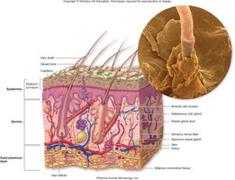
The skin, or integument, forms the primary boundary between the human body and the environment. It is composed of the skin itself, hair, nails, and associated glands. The skin's surface area ranges from 1.5 to 2 square meters, with thickness varying by location. The skin is organized into three main layers: the epidermis, dermis, and subcutaneous tissue.
Epidermis: Outermost layer, includes the stratum corneum (dead, keratinized cells) and stratum basale (source of new cells).
Dermis: Connective tissue layer containing fibroblasts, collagen, nerves, blood vessels, and glands.
Subcutaneous Layer: Adipose connective tissue providing insulation and energy storage.
Defenses of the Skin
The skin employs multiple defense mechanisms to prevent microbial colonization and infection:
Antimicrobial peptides: Positively charged molecules that disrupt bacterial membranes, keeping microbial counts low.
Sebum: Oily secretion with low pH and high lipid content; inhibits non-adapted microbes and supports normal microbiota.
Sweat: High salt and low pH environment; contains lysozyme, which breaks down bacterial peptidoglycan.
Physical removal: Sloughing of stratum corneum removes attached microorganisms daily.
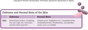
Normal Biota of the Skin
The skin is colonized by a diverse microbiota adapted to dry, salty, and acidic conditions. These organisms are sparsely distributed on dry areas but grow densely in moist regions and hair follicles. The Human Microbiome Project has revealed hundreds of microbial species inhabiting different skin layers, with individual variation and relative stability over time.
Common genera: Staphylococcus, Streptococcus, Corynebacterium, Propionibacterium, Pseudomonas, Lactobacillus, and yeasts such as Candida.
Major Bacterial Skin Infections
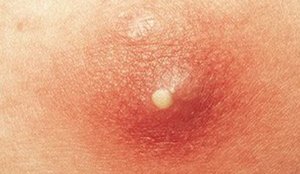
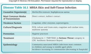
MRSA Skin and Soft Tissue Infection
Methicillin-resistant Staphylococcus aureus (MRSA) is a common cause of skin lesions, especially outside hospital settings. It is notable for its resistance to multiple antibiotics and its ability to survive harsh environmental conditions.
Signs and Symptoms: Raised, red, tender, pus-filled lesions, often localized around hair follicles; may be accompanied by fever.
Transmission: Direct or indirect contact with contaminated surfaces (e.g., gym equipment, razors).
Virulence Factors: Coagulase, hyaluronidase, staphylokinase, DNase, lipase.
Diagnosis: PCR, culture on blood or mannitol salt agar, catalase and coagulase tests.
Treatment: Incision and drainage, combination antibiotic therapy (e.g., clindamycin + TMP/SMZ, doxycycline).
Prevention: Good hygiene practices.
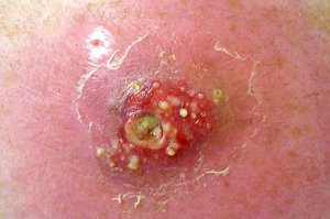
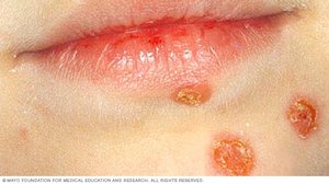
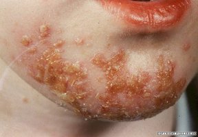
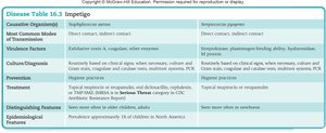
Impetigo
Impetigo is a superficial bacterial infection, most common in children aged 2 to 5, characterized by flaking or peeling skin and honey-colored crusts. It is caused by Staphylococcus aureus, Streptococcus pyogenes, or both.
Signs and Symptoms: Peeling skin, crusty/flaky scabs, honey-colored crusts, usually around the mouth, face, and extremities.
Pathogenesis: S. aureus produces exfoliative toxins; S. pyogenes is beta-hemolytic and can cause poststreptococcal complications.
Transmission: Direct or indirect contact with infected skin.
Treatment: Topical or oral antibiotics; hygiene is important for prevention.
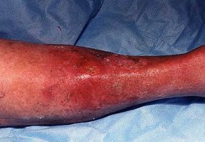
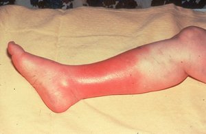
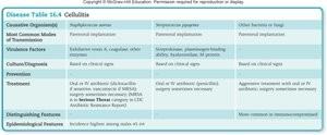
Cellulitis
Cellulitis is a fast-spreading infection of the dermis and subcutaneous tissues, most often caused by Staphylococcus aureus or Streptococcus pyogenes. It can occur in healthy individuals or those who are immunocompromised.
Signs and Symptoms: Red, swollen, tender skin; fever; lymphangitis (red streaks); possible bacteremia.
Transmission: Follows trauma or subtle skin breaks; can be caused by various bacteria or fungi in immunocompromised hosts.
Treatment: Oral or IV antibiotics; surgical debridement for severe cases.
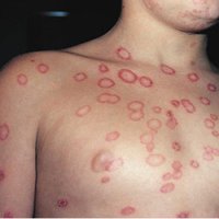
Ringworm (Dermatophytosis)
Ringworm refers to a group of fungal infections (dermatophytoses) affecting the nonliving epidermal tissues, hair, and nails. The causative agents are dermatophytes such as Trichophyton, Microsporum, and Epidermophyton.
Signs and Symptoms: Circular, red, scaly patches on the skin.
Diagnosis: UV fluorescence of infected hairs; KOH preparation of skin scrapings reveals fungal hyphae.
Treatment: Topical antifungals (azoles, terbinafine); therapy may last several weeks.
Maculopapular Rash Diseases
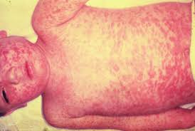
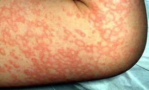
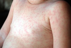
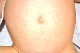
Measles (Rubeola)
Measles is a highly contagious viral disease caused by the measles virus (genus Morbillivirus). It is characterized by a red, maculopapular rash and systemic symptoms.
Transmission: Respiratory droplets; humans are the only reservoir.
Signs and Symptoms: Sore throat, cough, conjunctivitis, fever, rash starting on the head and spreading to the trunk and limbs.
Complications: Pneumonia, encephalitis, subacute sclerosing panencephalitis.
Prevention: MMR vaccine (live attenuated virus).
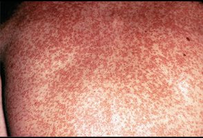
Rubella (German Measles)
Rubella is a mild viral rash disease caused by Rubivirus. It is of particular concern in pregnant women due to its teratogenic effects.
Signs and Symptoms: Pink macules and papules, joint pain in adults, teratogenic effects in fetuses (deafness, cardiac and ocular defects).
Transmission: Respiratory secretions; endemic worldwide.
Prevention: MMR vaccine.
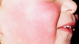
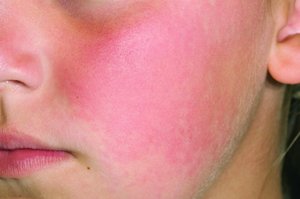
Fifth Disease (Erythema Infectiosum)
Fifth disease is a mild, contagious viral infection caused by parvovirus B19, notable for its "slapped cheek" rash in children.
Signs and Symptoms: Red cheeks, rash spreading to arms, legs, and trunk; may recur with heat or stress.
Transmission: Respiratory droplets; very contagious.
Treatment: No vaccine or specific therapy; disease is usually mild and self-limiting.
Roseola
Roseola, or "sixth disease," is a common viral infection in young children caused by human herpesvirus 6 (HHV-6) or HHV-7. It is characterized by a high fever followed by a maculopapular rash.
Signs and Symptoms: Sudden high fever, possible seizures, rash appearing as fever subsides.
Transmission: Nearly universal infection by adulthood; spread is not fully understood.
Treatment: Supportive care; no vaccine available.
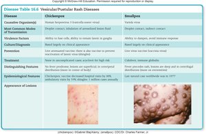
Vesicular and Pustular Rash Diseases
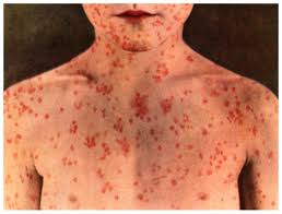
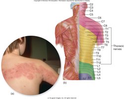
Chickenpox and Shingles (Varicella-Zoster Virus, HHV-3)
Chickenpox is a common, usually benign viral disease caused by human herpesvirus 3 (varicella-zoster virus). Shingles is a reactivation of the same virus, typically in older adults.
Chickenpox: Fever, centripetal vesicular rash, lesions at various stages of healing.
Shingles: Painful, localized vesicular rash following a dermatome; postherpetic neuralgia may occur.
Transmission: Respiratory droplets and lesion fluid; highly contagious.
Prevention: Live attenuated vaccine for chickenpox; Zostavax for shingles.
Treatment: Supportive care; acyclovir for at-risk patients.
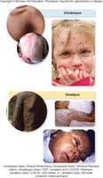
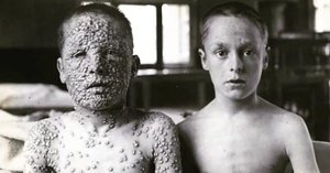
Smallpox (Variola Virus)
Smallpox is a highly contagious, often deadly viral disease caused by the variola virus. It has been eradicated worldwide through vaccination.
Signs and Symptoms: Fever, malaise, rash beginning in the mouth and spreading to the body, pustular lesions, scarring.
Transmission: Respiratory droplets, fomites (bedding, clothing).
Prevention: Vaccination (no longer routine due to eradication).
Treatment: None; supportive care only.
Other Skin Infections
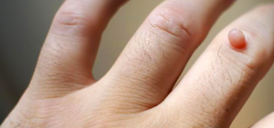
Warts (Human Papillomavirus, HPV)
Warts are benign skin growths caused by infection with human papillomavirus (HPV). They are most common on hands and feet and are transmitted by direct contact.
Types: Common warts, plantar warts, flat warts, and others depending on location and appearance.
Treatment: Removal by cryotherapy, salicylic acid, or other methods; many resolve spontaneously.
Leishmaniasis
Leishmaniasis is a protozoan infection transmitted by female sand flies, causing cutaneous or mucocutaneous lesions. It is endemic to tropical and subtropical regions.
Causative Agents: Leishmania tropica (cutaneous), Leishmania siliensis (mucocutaneous).
Transmission: Sand fly bites; zoonotic cycle.
Treatment: No vaccine; prevention relies on avoiding sand fly exposure.
Cutaneous Anthrax
Cutaneous anthrax is caused by Bacillus anthracis and results from endospore entry through skin abrasions. It is characterized by a painless, black eschar.
Transmission: Contact with infected animal hides or contaminated materials.
Treatment: Antibiotics; vaccine available for high-risk groups.
