 Back
BackPigments and Lipids: Histochemistry and Pathological Significance
Study Guide - Smart Notes
 Tailored notes based on your materials, expanded with key definitions, examples, and context.
Pigments and Lipids in Histochemistry
Overview
This study guide covers the classification, identification, and pathological significance of pigments and lipids in tissues, with a focus on histochemical techniques. Understanding these substances is crucial for diagnosing various diseases and interpreting tissue pathology in microbiology and pathology.
Pigments: Classification and Examples
General Classification of Pigments
Artefact Pigments: Deposits formed due to chemical reactions during tissue processing (e.g., formalin pigment, malarial pigment, mercury pigment, dichromate deposits).
Endogenous Pigments: Produced within tissues, either as part of normal physiology or as metabolic by-products (e.g., bile pigments, lipofuscin, melanin, iron, calcium, copper, uric acid/urates).
Exogenous Pigments: Enter the body from the environment, often with no physiological function (e.g., carbon, silica, asbestos).
Artefact Pigments
Formalin Pigment: Brown/black deposit after fixation in acid formalin, especially in hemorrhagic tissue. Removed with picric acid.
Malarial Pigment: Similar to formalin pigment, formed in/near RBCs with malaria parasite.
Mercury Pigment: Black deposit from mercury-containing fixatives. Removed with iodine and sodium thiosulphate.
Dichromate Deposits: Yellow/brown deposits after potassium dichromate fixation; removed with acid alcohol.
Endogenous Pigments
Bile Pigments: Products of RBC breakdown (bilirubin: red/brown; biliverdin: green). Accumulate in liver diseases or hemolytic conditions, leading to jaundice and cholestasis.
Lipofuscin: 'Wear and tear' pigment from lipid oxidation, seen near the nucleus. Stained by Sudan black, PAS, Schorl’s, Long Ziehl-Neelsen. Found in heart, liver, brain.
Melanin: Black/brown pigment produced by melanocytes. Found in skin, eye, hair, brain, and melanomas. Demonstrated by Masson Fontana stain.
Iron (Haemosiderin): Iron-storage complex, brown pigment, mainly from erythrocyte breakdown. Demonstrated by Perls’ Prussian Blue reaction.
Calcium: Absorbed from the gut, deposited in bone/teeth (hydroxyapatite), and in pathological calcification. Demonstrated by Von Kossa (black) and Alizarin (red) stains.
Copper: Essential for metabolism; accumulates in Wilson’s disease. Demonstrated by Shikata Orcein, Rubeanic acid, rhodanine stains.
Uric Acid/Urates: Breakdown products of purine nucleotides; form crystals in joints (gout). Crystals are birefringent under polarising microscopy.
Exogenous Pigments
Carbon: Most common exogenous pigment, seen in lungs of urban dwellers and smokers. Phagocytosed by macrophages.
Silica: Inert, angular masses in lungs/lymph nodes; may be birefringent. Causes silicosis.
Asbestos: Long, beaded fibers causing fibrosis and asbestosis. Asbestos bodies are coated with protein and haemosiderin, demonstrated by Perls’ stain.
Pathological Accumulation of Pigments
Haemosiderin, Haemochromatosis, and Haemosiderosis
Haemosiderin: Iron-storage complex in tissues, poorly available for metabolic needs. Seen in conditions with increased RBC breakdown.
Haemochromatosis: Inherited disorder with excessive iron absorption and deposition in parenchymal organs (liver, pancreas, heart). Leads to cirrhosis, diabetes, skin pigmentation, and organ failure. Treated with phlebotomy.
Haemosiderosis: Secondary iron overload, often due to repeated transfusions or chronic hemolysis. Iron stored mainly in reticuloendothelial cells; less toxic than haemochromatosis. Treated with iron chelators.
Histochemical Staining Methods
Perls’ Prussian Blue: Detects ferric iron (Fe3+) in tissues. Iron reacts with potassium ferrocyanide in acidic conditions to form blue pigment.
Masson Fontana: Demonstrates melanin as black granules.
Von Kossa: Silver impregnation method for calcium (black deposits).
Alizarin Red: Stains calcium deposits red.
Microscopy Techniques
Polarising Microscopy and Birefringence
Polarising microscopy is used to detect birefringent materials, such as urate crystals in gout. Birefringence is the property of a material to split light into two rays, making certain crystals visible against a dark background.
Lipids: Classification, Identification, and Pathology
Definition and Classification
Lipids: Hydrophobic molecules defined by their solubility in fat solvents and insolubility in water.
Conjugated Lipids: Neutral fats, waxes, cholesterol esters, phosphoglycerides, sphingomyelins, ceramides, glycolipids.
Unconjugated Lipids: Fatty acids, steroids.
Histochemical Identification of Lipids
Lysochrome Methods: Use lipid-soluble dyes (e.g., Sudan I-IV, Sudan Black, Oil Red O, Nile Blue) to stain lipids in frozen sections. Dyes are soluble in lipids but not in water, resulting in selective staining.
Osmium Tetroxide: Fixes and blackens lipids, but is rarely used in routine histochemistry.
Bright-field Microscopy: Used with H&E, Sudan-type stains, and Oil Red O for lipid visualization.
Polarised Light Microscopy: Used to detect birefringent lipid bodies (e.g., oval fat bodies in urine).
Fatty Liver Disease and Lipid Storage Disorders
Fatty Liver Disease: Accumulation of neutral fats in hepatocytes, often due to alcohol abuse or metabolic syndrome. Identified by Oil Red O or Sudan Black staining in frozen sections.
Lipid Storage Disorders (Lipidoses): Inherited metabolic diseases causing lipid accumulation in various organs, especially the CNS (e.g., Tay-Sachs, Fabry disease). Lead to neurodegeneration and organ dysfunction.
Summary Table: Pigment Types and Staining Methods
Pigment | Origin | Color | Staining Method | Pathological Significance |
|---|---|---|---|---|
Formalin pigment | Artefact | Brown/black | Removed with picric acid | Fixation artifact |
Bile pigments | Endogenous | Red/brown, green | H&E, special stains | Liver disease, jaundice |
Lipofuscin | Endogenous | Brown | Sudan black, PAS | Aging, neurodegeneration |
Melanin | Endogenous | Black/brown | Masson Fontana | Melanoma, pigmentation |
Haemosiderin | Endogenous | Brown | Perls’ Prussian Blue | Iron overload |
Calcium | Endogenous | Black (Von Kossa), red (Alizarin) | Von Kossa, Alizarin Red | Calcification, bone |
Carbon | Exogenous | Black | None (site identification) | Anthracosis, pneumoconiosis |
Asbestos | Exogenous | Brown (coated fibers) | Perls’ Prussian Blue | Asbestosis, mesothelioma |
Key Definitions
Birefringence: The property of a material to split light into two rays, detectable by polarising microscopy.
Neutral Fat: Triglycerides, the main storage form of fat in the body.
Lysochrome Principle: Staining based on the solubility of dyes in lipids, not on chemical reactions.
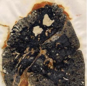
Example: Anthracosis (Coal Worker's Lung)
Anthracosis is the accumulation of carbon pigment in the lungs, commonly seen in urban dwellers and coal workers. The pigment is phagocytosed by macrophages and can be identified by its black color in lung tissue sections.
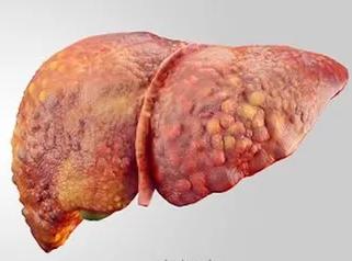
Example: Cirrhosis in Haemochromatosis
Haemochromatosis leads to excessive iron deposition in the liver, resulting in cirrhosis. The liver appears nodular and fibrotic, with a characteristic bronze discoloration due to iron overload.
Example: Perls’ Prussian Blue Staining
Perls’ Prussian Blue is a classic histochemical stain for detecting ferric iron in tissues. The reaction produces a blue pigment where iron is present, allowing for the identification of haemosiderin deposits in organs such as the liver, spleen, and heart.
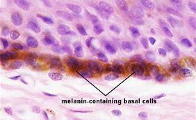
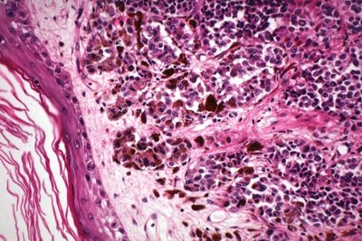
Example: Melanin in Skin
Melanin is a brown-black pigment produced by melanocytes in the basal layer of the epidermis. It provides protection against ultraviolet radiation and is increased in conditions such as melanoma.
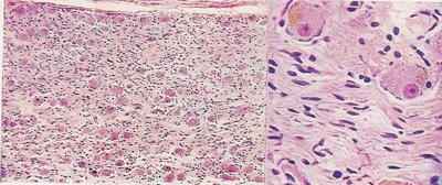
Example: Lipofuscin in Neurons
Lipofuscin appears as a light brown pigment in the cytoplasm of ganglion cells, especially in aging or neurodegenerative conditions. It is a marker of oxidative stress and cellular aging.
Example: Melanoma with Melanin-Laden Macrophages
In melanoma, melanin pigment can be found within macrophages and lymphocytes, indicating tumor invasion and immune response.
Example: Haemosiderin in Kidney
Haemosiderin deposits in the kidney are a result of hemoglobin breakdown and can be visualized using Perls’ Prussian Blue stain, indicating iron overload or chronic hemolysis.
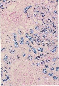
Example: Heart Failure Cells
Chronic congestion of the lung leads to the presence of 'heart failure cells'—macrophages containing haemosiderin, visible with H&E and Perls’ stains.
Example: Fatty Liver Disease
Alcohol-induced fatty liver disease is characterized by the accumulation of neutral fats in hepatocytes, which can be demonstrated by Oil Red O or Sudan Black staining in frozen sections.
Example: Lipid Storage Disorders
Lipid storage disorders, such as Fabry disease and Tay-Sachs disease, result in the accumulation of lipids in various tissues, particularly affecting the nervous system and kidneys.
Additional info: This guide integrates histochemical principles with clinical pathology, providing a comprehensive overview for microbiology and pathology students.
