 Back
BackPreparation and Staining of Bacterial Smears: Techniques and Applications
Study Guide - Smart Notes
 Tailored notes based on your materials, expanded with key definitions, examples, and context.
Preparation and Staining of Bacterial Smears
Introduction
Staining and preparation of bacterial smears are foundational techniques in microbiology, enabling visualization of microbial morphology and cellular structures. These methods enhance contrast and allow for differentiation between types of bacteria, which is essential for identification and study.
Preparation of Smears
Principles and Rationale
Smear Preparation: Involves spreading a thin film of bacterial cells on a clean glass slide, followed by air-drying and fixation.
Purpose: Fixation kills bacteria, adheres them to the slide, and preserves cellular morphology for staining.
Fixation Methods: Most commonly by heat (passing through a flame) or chemically (e.g., 95% methanol for 1 minute).
Autolysis Prevention: Fixation denatures bacterial enzymes, preventing self-digestion (autolysis).
Procedures for Preparing Smears
From Solid Media: Place 1–2 loopfuls of water on the slide, mix with a small amount of culture, and air-dry.
From Liquid Media: Place 2–3 loopfuls of broth culture directly on the slide and air-dry.
Fixation: Pass the dried slide through a flame 2–3 times or cover with methanol for 1 minute.
Cleaning: Slides must be grease-free for proper smear adherence.
Why Wait Until Smears Are Dry Before Fixing?
Preserving Cell Morphology: Heat-fixing wet smears can cause cells to rupture and distort.
Ensuring Proper Adhesion: Wet smears may wash off during staining, leading to sample loss.
Examples of Bacteria Used in Smear Preparation
Escherichia coli (Gram-negative rod): Forms cream-colored colonies on nutrient agar.
Staphylococcus aureus (Gram-positive cocci): Forms golden-yellow colonies on solid media.
Staining Techniques
Purpose of Staining
Enhances contrast between bacteria and background for better visualization.
Allows observation of cellular details and differentiation between bacterial types.
Types of Staining Techniques
Simple Staining: Uses a single reagent; all bacteria are stained similarly.
Differential Staining: Uses multiple reagents; bacteria react differently (e.g., Gram staining).
Structural Staining: Identifies specific parts of microorganisms (e.g., flagella, spores).
Simple Staining
Direct Stain: Stains the bacteria directly (e.g., methylene blue, crystal violet, safranin).
Negative Stain: Stains the background, leaving bacteria clear (e.g., nigrosin, eosin).
Stain Chemistry: Most stains are synthetic aniline dyes, often salts with charged chromophores.
Basic Stains: Positively charged, attracted to negatively charged cell components.
Acidic Stains: Negatively charged, repelled by bacterial surfaces, stain the background.
Simple Staining Procedure
Hold slide with forceps or a rack.
Cover smear with methylene blue for 30–60 seconds.
Rinse with distilled water and blot dry.
Examine under microscope using oil immersion if needed.
Negative Staining
Bacteria appear clear/bright against a dark background.
Uses colloidal stains (e.g., India ink, nigrosin) that do not penetrate cells.
No heat-fixing or strong chemicals are used, preserving cell morphology.
Especially useful for visualizing coccobacilli and other delicate bacteria.
Negative Staining Procedure
Place a drop of nigrosin at the end of a clean slide.
Mix with a small amount of culture (add water if from solid media).
Spread the drop with another slide to create a gradient smear.
Allow to air-dry; do not heat-fix.
Gram Staining
Principle and Importance
The Gram stain is a differential staining technique that classifies bacteria as Gram-positive or Gram-negative based on cell wall structure. Developed by Hans Christian Gram in 1884, it remains a cornerstone in bacterial identification and clinical diagnostics.
Gram Staining Procedure
Apply primary stain (crystal violet) – all cells stain purple.
Add mordant (Gram’s iodine) – forms a crystal violet-iodine complex (CV-I).
Decolorize with ethanol – removes stain from Gram-negative cells only.
Counterstain with safranin – stains decolorized cells pink/red.
Mechanism and Interpretation
Gram-positive bacteria: Thick peptidoglycan layer retains CV-I complex; cells appear purple.
Gram-negative bacteria: Thin peptidoglycan and outer membrane; CV-I complex is washed out, cells take up safranin and appear pink/red.
Cell Wall Structure: Gram-positive: multiple peptidoglycan layers; Gram-negative: thin peptidoglycan, outer membrane with lipopolysaccharides.
Clinical Relevance
Most Gram-negative bacteria are pathogenic and more resistant to antibiotics.
Gram-positive bacteria are often non-pathogenic and more susceptible to antibiotics.
Staining Step | Gram-Positive | Gram-Negative |
|---|---|---|
Crystal Violet | Purple | Purple |
Gram's Iodine | Purple (CV-I complex) | Purple (CV-I complex) |
Decolorizer (Ethanol) | Purple (retained) | Colorless (CV-I lost) |
Safranin | Purple | Pink/Red |
Summary Table of Stains
Stain Type | Purpose | Examples |
|---|---|---|
Simple Stain | Visualize cell shape and arrangement | Methylene blue, Crystal violet |
Negative Stain | Visualize capsules, delicate bacteria | Nigrosin, India ink |
Gram Stain | Differentiates Gram-positive/negative | Crystal violet, Safranin |
Key Terms and Definitions
Smear: Thin film of bacteria on a slide for microscopic examination.
Fixation: Process of adhering and killing bacteria on a slide.
Chromophore: The colored ion in a dye responsible for its color.
Basic Stain: Positively charged dye that binds to negatively charged cell components.
Acidic Stain: Negatively charged dye that stains the background.
Mordant: Substance that stabilizes dye binding (e.g., iodine in Gram stain).
Decolorizer: Removes primary stain from certain cells (e.g., ethanol in Gram stain).
Counterstain: Secondary stain that colors decolorized cells.
References to Images
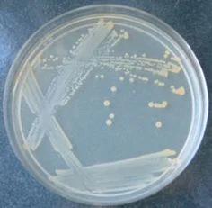
- : Example of bacterial growth on solid media (e.g., E. coli or S. aureus).
Additional info: The above notes integrate foundational microbiology laboratory techniques, including the rationale, procedures, and clinical relevance of bacterial smear preparation and staining. These concepts are essential for understanding microbial identification and classification in both research and clinical settings.
