 Back
BackPrinciples and Applications of Microscopy and Staining in Microbiology
Study Guide - Smart Notes
 Tailored notes based on your materials, expanded with key definitions, examples, and context.
Microscopy in Microbiology
Principles of Light Microscopy
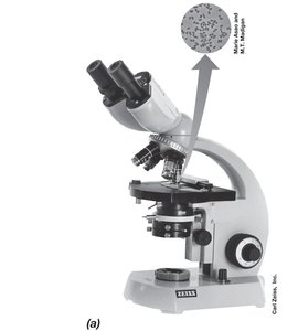
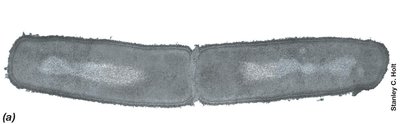
Light microscopy is a fundamental technique in microbiology, allowing visualization of cells and microorganisms using visible light. The compound light microscope is the most common instrument, utilizing two sets of lenses (objective and ocular) to magnify specimens.
Bright-field microscopy: Specimens are visualized due to differences in contrast (density) between the specimen and its surroundings.
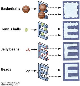
Magnification: The ability to enlarge an object. Total magnification is calculated as:
Maximum magnification: Approximately 2,000× for light microscopes.
Resolution: The ability to distinguish two adjacent objects as separate and distinct. Determined by the wavelength of light and the numerical aperture (NA) of the lens.
Limit of resolution: About 0.2 μm for light microscopes.

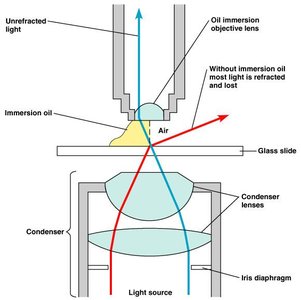
Refractive Index and Immersion Oil
The refractive index is the light-bending ability of a medium. Immersion oil is used with high-magnification lenses to prevent light from bending away from the lens, thereby improving resolution.
Immersion oil: Matches the refractive index of glass, minimizing light loss.
Without immersion oil: Most light is refracted and lost, reducing image clarity.
Improving Contrast in Light Microscopy
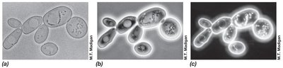
Phase-Contrast and Dark-Field Microscopy
Contrast enhancement techniques allow visualization of live, unstained specimens.
Phase-contrast microscopy: Uses a phase ring to amplify differences in refractive index, producing dark cells on a light background.
Dark-field microscopy: Illuminates specimens from the side; only scattered light enters the lens, resulting in light images on a dark background. Useful for observing motility.
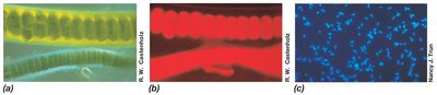
Fluorescence Microscopy
Fluorescence microscopy is used to visualize specimens that emit light of one color when illuminated with another color. Fluorochromes are dyes that fluoresce under UV light.
Autofluorescence: Some cells naturally fluoresce.
DAPI: A fluorescent dye used for staining DNA, widely applied in clinical diagnostics and microbial ecology.
Immunofluorescence: Uses fluorescent dyes linked to antibodies for specific identification of microorganisms.
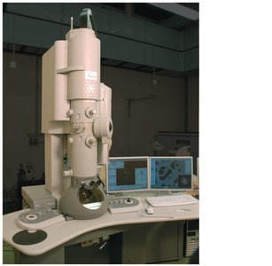
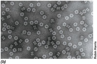
Electron Microscopy
Transmission and Scanning Electron Microscopy
Electron microscopes use electrons instead of photons, providing much higher resolution and magnification.
Transmission Electron Microscopy (TEM): Electromagnets act as lenses; operates in a vacuum. Allows visualization of structures at the molecular level. Specimens must be thin and stained.
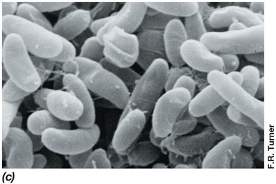
Scanning Electron Microscopy (SEM): Specimens are coated with heavy metals; an electron beam scans the surface, and scattered electrons are detected to produce a 3D image. Suitable for large specimens.
Magnification range: 15×–100,000× for SEM.
Advanced Microscopy Techniques
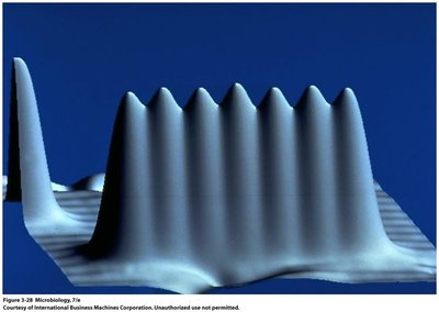
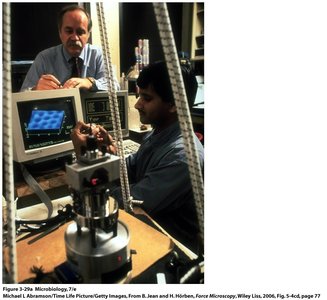
Scanning tunneling microscopes (STM) and atomic force microscopes (AFM) provide atomic-level resolution and 3D imaging.
STM: Uses a metal probe to scan surfaces at the atomic level.
AFM: Allows 3D imaging and measurement of forces at the molecular level.
Staining Techniques in Microbiology
Principles and Types of Staining
Staining improves contrast in microscopy, allowing better visualization and differentiation of cellular structures.
Dyes: Organic compounds that bind to specific cellular materials. Common stains include methylene blue, safranin, and crystal violet.
Stains: Consist of a positive and negative ion. Basic dyes have a cationic chromophore; acidic dyes have an anionic chromophore.
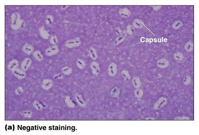
Negative staining: Stains the background instead of the cell.
Simple Stains
Simple staining uses a single dye to color cells, making them easier to see.
Examples: Methylene blue, crystal violet, eosin.
Mordant: A substance used to hold the stain or coat the specimen to enlarge it.
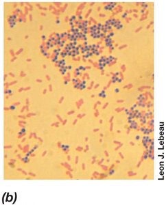
Differential Stains: The Gram Stain
The Gram stain is a differential staining technique that separates bacteria into two groups based on cell wall structure.
Gram-positive: Retain crystal violet and appear purple.
Gram-negative: Lose crystal violet, take up safranin, and appear red.
Procedure:
Flood heat-fixed smear with crystal violet (1 min).
Add iodine solution (1 min).
Decolorize with alcohol (20 sec).
Counterstain with safranin (1–2 min).
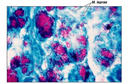
Differential Stains: Acid-Fast Stain
Acid-fast staining identifies cells that retain a basic stain in the presence of acid-alcohol, such as Mycobacterium species.
Acid-fast cells: Retain stain; non–acid-fast cells lose stain and are counterstained.
Special Stains
Special stains are used to highlight specific structures such as capsules, endospores, and flagella.
Negative staining: Useful for visualizing capsules.
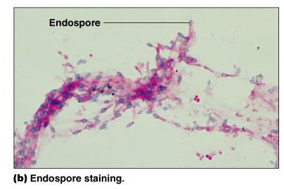
Endospore staining: Requires heat to drive the stain into spores.
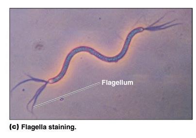
Flagella staining: Uses a mordant to make flagella visible.
Review of Staining Techniques
Staining techniques are classified as simple or differential. The choice of technique depends on the purpose (e.g., visualization, differentiation, identification), the reagents used, and the function of mordants in the process.
Simple stains: Use one dye; highlight entire cell.
Differential stains: Use multiple dyes; distinguish between cell types or structures.
Mordant: Enhances staining by binding dye to cell structures.
Staining Technique | Type | Purpose | Reagents | Mordant |
|---|---|---|---|---|
Simple Stain | Simple | Visualize cells | Methylene blue, crystal violet | May use mordant |
Gram Stain | Differential | Differentiate Gram-positive/negative | Crystal violet, iodine, alcohol, safranin | Iodine |
Acid-Fast Stain | Differential | Identify acid-fast bacteria | Carbol fuchsin, acid-alcohol, methylene blue | None |
Negative Stain | Special | Visualize capsules | India ink, nigrosin | None |
Endospore Stain | Special | Visualize endospores | Malachite green, heat, safranin | Heat |
Flagella Stain | Special | Visualize flagella | Carbol fuchsin, mordant | Mordant (e.g., tannic acid) |
Additional info: Staining techniques are essential for microbial identification, classification, and understanding cell structure and function. Proper selection and application of stains and microscopy methods are critical for accurate microbiological analysis.
