 Back
BackProkaryotes: Bacteria, Archaea, and Eukaryotes – Structure, Function, and Pathogenicity
Study Guide - Smart Notes
 Tailored notes based on your materials, expanded with key definitions, examples, and context.
Prokaryotes: Bacteria – Cell Structure and Classification
Genetic Relationships and the Tree of Life
Understanding the genetic relationships among all organisms is fundamental to microbiology. Scientists use universally conserved genes, such as the 16S rRNA gene, to build the "Tree of Life" and determine evolutionary ancestry.
Universally Conserved Genes: Essential genes present in all living cells, evolving slowly due to their critical function.
Molecular Phylogeny: DNA sequence alignment reveals evolutionary relationships; fewer differences indicate a recent common ancestor.
16S rRNA Gene: Contains both conserved and variable regions, allowing precise comparison across species.
Carl Woese's Discovery: Revealed Archaea as a distinct domain, splitting life into Bacteria, Archaea, and Eukarya.
Prokarya vs. Eukarya: Genetic and Structural Comparison
Prokaryotes (Bacteria and Archaea) differ from Eukaryotes in their cellular organization and genetic properties.
Bacteria: Circular DNA, 70S ribosomes, cell walls with peptidoglycan.
Archaea: Circular DNA, 70S ribosomes, transcription/translation machinery similar to Eukaryotes, cell walls without peptidoglycan.
Eukarya: Linear DNA, 80S ribosomes, complex endomembrane system, no peptidoglycan.
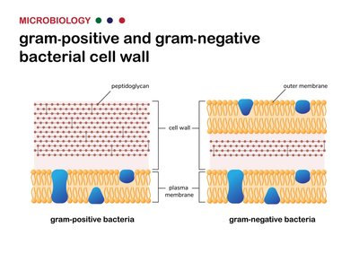
Cell Wall Morphology: Gram-Positive vs. Gram-Negative
The bacterial cell wall is a key structural feature, distinguishing Gram-positive from Gram-negative bacteria based on peptidoglycan thickness and the presence of an outer membrane.
Gram-Positive: Thick, multi-layered peptidoglycan reinforced with teichoic acids; retains Crystal Violet stain (purple).
Gram-Negative: Thin peptidoglycan layer, outer membrane with porins and lipopolysaccharide (LPS); does not retain Crystal Violet, appears pink/red.
Capsule: Organized polysaccharide layer outside the cell wall, prevents phagocytosis by immune cells.
Other Structural Features: Pili, Flagella, Nucleoid
Bacteria possess specialized structures for motility, adhesion, and genetic organization.
Pili (Fimbriae): Short, hair-like appendages involved in twitching motility and conjugation (DNA transfer).
Flagella: Long, helical filaments used for swimming, powered by the Proton Motive Force (PMF).
Nucleoid: Irregular region containing the bacterial chromosome, dense with DNA, RNA, and protein.
Cell Shape and Arrangement
Bacterial cell shape is determined by the cell wall and cytoskeletal proteins such as MreB.
Cocci: Spherical cells.
Bacilli: Rod-shaped cells.
Vibrio: Comma-shaped cells.
Spirochetes: Flexible, corkscrew-shaped cells.
Cell Growth, Metabolism, and Biofilms
Bacterial Growth Curve
Bacterial populations grow in distinct phases, each with unique metabolic characteristics.
Lag Phase: Cells adapt to environment, synthesize enzymes, no division.
Log (Exponential) Phase: Rapid, constant doubling; most susceptible to antibiotics.
Stationary Phase: Birth rate equals death rate; waste accumulates, pH drops.
Death Phase: Nutrients depleted, cells die exponentially.
Biofilms and Medical Implications
Biofilms are multicellular-like bacterial communities encased in extracellular polymeric substances (EPS), posing significant medical challenges.
EPS (Slime): Physical barrier protecting bacteria from immune cells and antibiotics.
Medical Issues: Biofilms on heart valves and catheters are resistant to immune clearance.
Bacterial Life Cycle Stages
Planktonic: Free-swimming individual cells.
Swarming: Coordinated group movement across surfaces.
Fruiting Bodies: Aggregated structures for spore production under starvation.
Sporulation
Bacteria form highly resistant spores under environmental stress, protecting DNA for extended periods.
Forespore: Asymmetrical division, coated with dipicolinic acid and calcium.
Bacterial Motility, Chemotaxis, and Adhesion
Flagellar Motility and Visualization
Bacterial flagella are complex machines enabling movement; in Spirochetes, flagella are internal (endoflagella).
Run and Tumble: Alternating forward movement (run) and random reorientation (tumble).
Types of Taxis
Chemotaxis: Movement toward or away from chemicals.
Aerotaxis: Movement toward oxygen.
Magnetotaxis: Alignment with Earth's magnetic field.
Phototaxis: Movement toward light.
Adhesion and Colonization
Attachment to surfaces is a critical step in infection, mediated by pili, fimbriae, and specialized adhesins.
Holdfasts: Extremely strong biological "glue" for surface attachment.
MSCRAMMs: Surface proteins binding to host matrix molecules (e.g., collagen).
E. coli Colonization: Two-step attachment: loose via Bundle Forming Pili, tight via injected Tir receptor.
Bacterial Pathogenicity: Extracellular and Intracellular Pathogens
Extracellular vs. Intracellular Pathogens
Bacterial pathogens are classified by their location during infection.
Extracellular: Multiply in blood/lymph, face antibodies and complement.
Intracellular: Multiply inside host cells, evade lysosomal destruction.
Examples and Disease Mechanisms
Extracellular: Propionibacterium acnes (acne), Helicobacter pylori (ulcers), Staphylococcus aureus (MRSA), Pseudomonas aeruginosa (biofilms in cystic fibrosis).
Intracellular: Listeria monocytogenes, Salmonella enterica, Mycobacterium tuberculosis, Chlamydia.
Host Survival Strategies
Spreading Factors: Enzymes (hyaluronidase, streptokinase) break down host barriers.
Nutrient Acquisition: Exoenzymes digest host tissues; iron acquisition via siderophores.
Granuloma Formation: Immune cells wall off bacteria (e.g., M. tuberculosis).
Intracellular Escape: Toxins (Listeriolysin O) allow escape from phagosomes.
Obligate Intracellular Life Cycle: Biphasic forms (Chlamydia: elementary and reticulate bodies).
Bacterial Toxins and Secretion Systems
Toxin Types and Effects
Endotoxins: Lipid A triggers cytokine release, causing septic shock.
Superantigens: Activate massive numbers of T-cells, causing cytokine storm.
Exotoxins: Membrane-disrupting (hemolysins), A-B toxins (diphtheria, cholera), metalloproteases (botulinum, tetanus).
Toxin Delivery Systems
Type 1-7 Secretion Systems: Specialized protein pumps for toxin export.
Type 3 Secretion System (T3SS): Molecular syringe injects toxins directly into host cells.
Archaea: Unique Features and Environmental Roles
Phylogenetic Relationships and Extremophiles
16S rRNA: Defines Archaea as a unique domain.
Crenarchaeota: Thermophiles.
Euryarchaeota: Methanogens, halophiles.
Cell Structure and Metabolism
S-layer: Protein armor instead of peptidoglycan.
Membrane Lipids: Ether-linked, branched, cyclized for stability.
Hami: Grappling hook-like fimbriae for attachment.
Methanogenesis: Conversion of CO2 and H2 to methane, crucial for global carbon cycling.
Eukaryotes: Algae, Fungi, and Protozoans
Cellular Features and Endosymbiosis
80S Ribosomes: Larger than prokaryotic ribosomes.
Nucleus: Membrane-enclosed chromosome.
Cilia: Microtubule-based motility structures unique to eukaryotes.
Endosymbiotic Theory: Mitochondria and chloroplasts originated from prokaryotes.
Algae: Diversity and Ecological Roles
Photoautotrophs: Major oxygen producers.
Pigments: Phycoerythrin (red), chlorophyll (green), cell wall components (agar, carrageenan, alginate).
Holdfasts: Anchoring structures, not roots.
Fungi: Structure, Function, and Pathogenicity
Chemoheterotrophs: Absorb nutrients from dead organisms.
Yeasts and Molds: Unicellular and multicellular forms; dimorphism as a virulence factor.
Cell Wall: Chitin and galactomannans.
Basidiomycetes: Mushroom life cycle, spore production.
Lichens: Mutualistic communities of fungi and algae/cyanobacteria.
Pathogenic Fungi
Antifungal Targets: Ergosterol in cell membrane.
Ascomycetes: Penicillium (antibiotics, cheese), Aspergillus (aflatoxins, ergot alkaloids).
Yeasts: Candida albicans (phenotypic switching), Cryptococcus neoformans (capsule, meningitis).
Dermatophytes: Trichophyton rubrum (athlete’s foot, ringworm).
Protozoan Parasites: Life Cycles and Disease
Life Stages and Motility
Trophozoite: Active, reproducing form inside hosts.
Cyst: Dormant, infectious form outside hosts; resistant to chlorine.
Motility: Cilia (Paramecium), pseudopods (Amoeba), flagella (Euglena).
Parasitic Diseases
Amoebas: Entamoeba histolytica (dysentery), Naegleria fowleri (encephalitis).
Flagellates: Giardia intestinalis (diarrhea), Trichomonas vaginalis (STD).
Vector-borne: Trypanosoma brucei (sleeping sickness), Leishmania (leishmaniasis).
Apicomplexa: Plasmodium falciparum (malaria), Toxoplasma gondii (congenital defects).
Viruses: Structure, Replication, and Disease
Virus Structure and Classification
Virion: Static particle outside host, composed of capsid and genetic material (DNA or RNA).
Envelope: Lipid membrane from host cell, fragile and easily disrupted.
Bacteriophage Life Cycles
Lytic Cycle: Attachment, entry, synthesis, assembly, release (host cell lysis).
Lysogenic Cycle: Integration as prophage, replication with host, induction to lytic cycle.
Animal Virus Replication
Attachment and Entry: Entire virus enters cell, uncoating releases genome.
DNA Viruses: Replicate in nucleus, use host machinery.
RNA Viruses: High mutation rates, four types (+ssRNA, -ssRNA, retroviruses, dsRNA).
Release: Nonenveloped viruses via lysis; enveloped viruses via budding.
Viral Diseases and Latency
DNA Viruses: Pox, Herpes, Papilloma, Adenovirus, Hepadnavirus.
RNA Viruses: Coronaviruses, influenza, HIV (retrovirus), rotavirus.
Latency: Herpes hides in nerve ganglia; HIV integrates permanently, leading to AIDS.
Oncoviruses: Linked to cancer, disrupt tumor suppression.
