 Back
Backlecture 10
Study Guide - Smart Notes
 Tailored notes based on your materials, expanded with key definitions, examples, and context.
Prokaryotic Diversity and Classification
The Tree of Life and Biological Classification
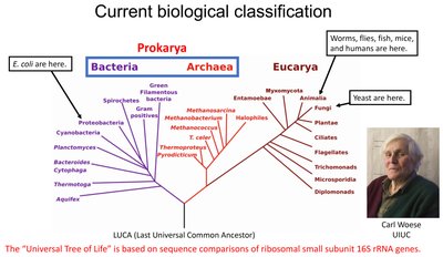
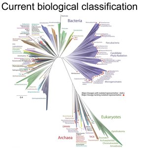
The classification of living organisms has evolved from the five-kingdom system to a three-domain system based on molecular data, particularly 16S rRNA gene sequences. This modern approach divides life into Bacteria, Archaea, and Eukarya, reflecting evolutionary relationships and genetic differences.
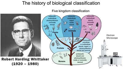
Five Kingdoms: Plants, Animals, Fungi, Protists, Monera (prokaryotes).
Three Domains: Bacteria (true bacteria), Archaea (ancient prokaryotes), Eukarya (organisms with a nucleus).
16S rRNA gene sequencing is the molecular basis for current classification, providing a universal marker for phylogenetic studies.
Bacterial Cell Structure and Morphology
Typical Prokaryotic Cell Structure
Prokaryotic cells, such as bacteria, have a simple internal structure but possess specialized features for survival and adaptation. Key components include:
Cell wall: Provides structural support and shape; composition differs between Gram-positive and Gram-negative bacteria.
Plasma membrane: Controls transport of substances in and out of the cell.
Capsule: A polysaccharide layer that protects against desiccation and immune attack.
Pili and fimbriae: Surface appendages for attachment and genetic exchange.
Flagella: Motility structures enabling movement.
Nucleoid: Region containing the bacterial chromosome (DNA).
Plasmids: Small, circular DNA molecules carrying accessory genes.
Ribosomes: Sites of protein synthesis.

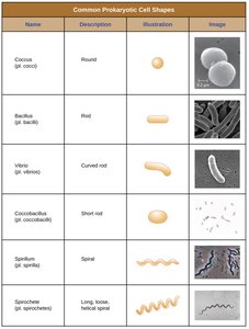
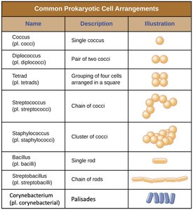
Prokaryotic Cell Shapes and Arrangements
Bacteria exhibit a variety of shapes and arrangements, which are important for identification and classification.
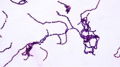
Shapes: Cocci (spherical), bacilli (rod-shaped), vibrios (curved rods), spirilla (spiral), spirochetes (flexible spirals), coccobacilli (short rods).
Arrangements: Single, pairs (diplococci), chains (streptococci), clusters (staphylococci), tetrads, palisades.
Name | Description | Illustration | Image |
|---|---|---|---|
Coccus | Round | ● | Micrograph of cocci |
Bacillus | Rod | ▬ | Micrograph of bacilli |
Vibrio | Curved rod | ~ | Micrograph of vibrio |
Coccobacillus | Short rod | ◦ | Micrograph of coccobacilli |
Spirillum | Spiral | ∿ | Micrograph of spirilla |
Spirochete | Long, loose helical spiral | ∿∿ | Micrograph of spirochetes |
Name | Description | Illustration |
|---|---|---|
Coccus | Single coccus | ● |
Diplococcus | Pair of two cocci | ●● |
Tetrad | Grouping of four cells in a square | ●● ●● |
Streptococcus | Chain of cocci | ●●●●● |
Staphylococcus | Cluster of cocci | ●●● ●●● |
Bacillus | Single rod | ▬ |
Streptobacillus | Chain of rods | ▬▬▬▬ |
Corynebacterium | Palisades | ||||| |
Complex Life Cycles of Bacteria
Bacterial Growth and Life Cycle Phases
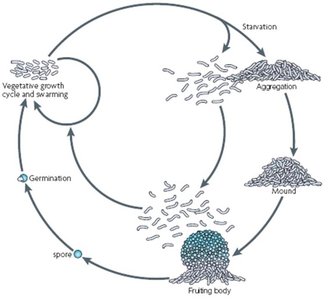
Bacteria can exist in different physiological states and undergo complex life cycles, especially in response to environmental changes.
Planktonic phase: Free-swimming, motile cells.
Swarming phase: Coordinated movement across surfaces.
Attachment and aggregation: Cells adhere to surfaces and to each other, forming microcolonies.
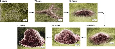
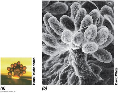
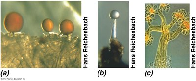
Fruiting body formation: Under starvation, some bacteria aggregate into multicellular structures (e.g., myxobacteria).
Sporulation: Formation of resistant spores or endospores for survival under harsh conditions.
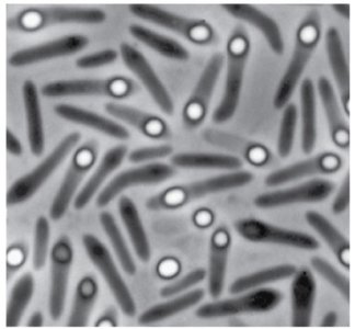
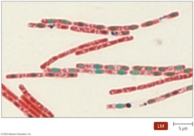
Endospore Formation and Visualization
Some Gram-positive bacteria, such as Bacillus and Clostridium, form endospores to survive extreme conditions. Endospores are highly resistant to heat, desiccation, UV light, and chemicals.
Schaeffer-Fulton stain: A differential staining technique used to visualize endospores (endospores appear green, vegetative cells red).
Medical relevance: Endospore-forming bacteria can cause persistent infections and are difficult to eradicate.
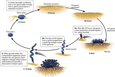
Myxobacteria and Actinomycetes: Complex Sporulation
Some bacteria, such as myxobacteria and actinomycetes, exhibit multicellular behaviors and complex sporulation cycles.
Myxobacteria: Form fruiting bodies and spores under starvation; display social behavior and coordinated movement.
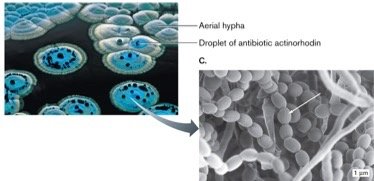
Actinomycetes: Gram-positive, filamentous bacteria that form spores and are often mistaken for fungi; important for antibiotic production.
Biofilms: Structure, Function, and Medical Relevance
Biofilm Formation and Properties
Biofilms are structured communities of bacteria encased in a self-produced extracellular matrix, often attached to surfaces. Biofilm development involves several stages:
Initial attachment: Free-floating (planktonic) cells adhere to a surface.
Matrix production: Secretion of extracellular polysaccharides forms a protective matrix.
Community maturation: Cells divide, communicate, and differentiate within the biofilm.
Dispersion: Some cells return to the planktonic state to colonize new sites.
Biofilms provide increased resistance to antibiotics, protection from environmental stress, and enable metabolic cooperation among cells.
Medical and Environmental Impact of Biofilms
Biofilms are implicated in a variety of medical and environmental contexts:
Medical devices: Catheters, implants, and prosthetics are common sites for biofilm-associated infections.
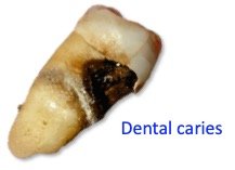
Chronic infections: Pseudomonas aeruginosa in cystic fibrosis lungs, dental plaque leading to caries and periodontal disease.
Environmental biofilms: Found on rocks, in waterways, and on marine snow (aggregates of organic material in oceans).
Antibiotic resistance: Biofilm bacteria are much more resistant to antimicrobial agents than planktonic cells.
Biofilm Detection and Control
Biofilm formation can be detected by staining with crystal violet, which binds to extracellular polysaccharides. Control strategies include physical removal, chemical disinfection, and filtration.
Crystal violet assay: Quantifies biofilm biomass by staining the matrix.
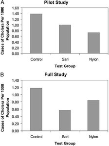
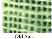
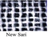
Filtration: Simple filtration methods, such as using folded sari cloth, can reduce waterborne diseases like cholera by trapping bacteria in biofilms.

Summary Table: Key Features of Prokaryotic Life Cycles and Biofilms
Feature | Description | Example |
|---|---|---|
Planktonic phase | Free-swimming, motile cells | Vibrio cholerae |
Biofilm | Community of cells in extracellular matrix | Dental plaque |
Endospore | Dormant, resistant cell type | Bacillus anthracis |
Fruiting body | Multicellular structure for spore dispersal | Myxobacteria |
Additional info: The study of prokaryotic diversity, structure, and life cycles is foundational for understanding microbial ecology, pathogenesis, and strategies for controlling infectious diseases.
