Back
BackProkaryotic Cell Structure and Function: A Study Guide
Study Guide - Smart Notes
 Tailored notes based on your materials, expanded with key definitions, examples, and context.
Prokaryotic Cell Basics
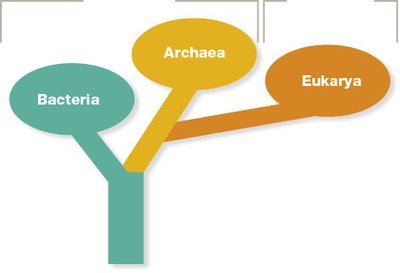
Domains of Prokaryotes
Prokaryotic cells are classified into two main domains: Bacteria and Archaea. These domains are distinct from the domain Eukarya, which includes all eukaryotic organisms.
Similarity: Both Bacteria and Archaea lack a membrane-bound nucleus and membrane-bound organelles.
Difference: Bacteria have cell walls containing peptidoglycan, while Archaea have pseudopeptidoglycan or other unique cell wall components.
Basic Description of Prokaryotic Cells
Prokaryotic cells are generally small (0.2–2.0 μm in diameter), which allows for efficient nutrient uptake and waste removal due to a high surface-area-to-volume ratio. They lack a true nucleus, and their genetic material is located in a region called the nucleoid.
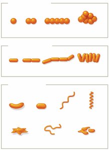
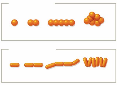
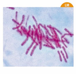
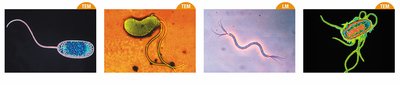
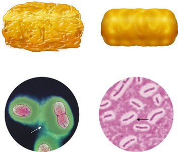
Shapes and Arrangements of Prokaryotes
Prokaryotes exhibit a variety of shapes and arrangements, which are important for identification and classification.
Bacilli: Rod-shaped (singular: Bacillus)
Cocci: Spherical (singular: Coccus)
Other shapes: Vibrio (comma-shaped), Spirillum (spiral), Spirochete (flexible spiral), Coccobacillus (short rod), Stella (star-shaped), Filamentous, Pleomorphic (variable shape)
Arrangements result from cell division patterns:
Diplo-: Pairs
Strepto-: Chains
Staphylo-: Clusters (mainly cocci)
Palisades: Side-by-side arrangement (mainly bacilli)
Pleomorphism
Pleomorphic organisms can alter their shape or size in response to environmental conditions. This property can enhance their ability to evade the immune system or adapt to different environments, impacting their pathogenicity.
Binary Fission
Prokaryotic cells reproduce asexually by binary fission:
DNA is replicated.
The cell elongates, and chromosomes are segregated to opposite ends.
A septum forms at the midpoint, dividing the cell into two genetically identical daughter cells.
Additional info: Binary fission is a rapid process, allowing prokaryotes to multiply quickly under favorable conditions.
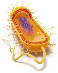
Extracellular Structures of Prokaryotes
Plasma Membrane
The plasma membrane is a thin, flexible phospholipid bilayer with embedded proteins, acting as a selective barrier. Its fluidity is influenced by temperature and fatty acid composition:
Bacteria: Linear fatty acids
Archaea: Long-branched fatty acids; some form monolayers for stability in extreme environments
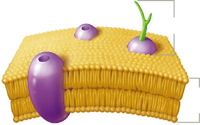
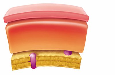
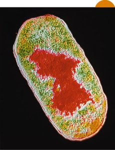
Cell Wall
The cell wall provides rigidity and protection:
Bacteria: Peptidoglycan
Archaea: Pseudopeptidoglycan or other polymers
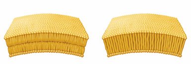
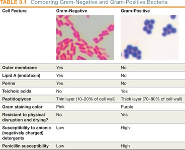
There are two main types of bacterial cell walls:
Gram-positive: Thick peptidoglycan layer, teichoic acids, no outer membrane
Gram-negative: Thin peptidoglycan layer, outer membrane with lipopolysaccharide (LPS), porins
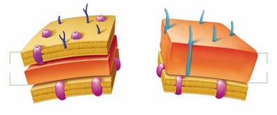
Feature | Gram-Negative | Gram-Positive |
|---|---|---|
Outer membrane | Yes | No |
Lipid A (endotoxin) | Yes | No |
Porins | Yes | No |
Teichoic acids | No | Yes |
Peptidoglycan | Thin (10–20%) | Thick (70–80%) |
Gram stain color | Pink | Purple |
Physical resistance | Low | High |
Penicillin susceptibility | Low | High |
Acid-Fast Bacteria
Acid-fast bacteria, such as Mycobacterium and Nocardia, have a waxy mycolic acid layer in their cell walls. Acid-fast staining is clinically useful for identifying these pathogens, which are resistant to desiccation and many disinfectants.
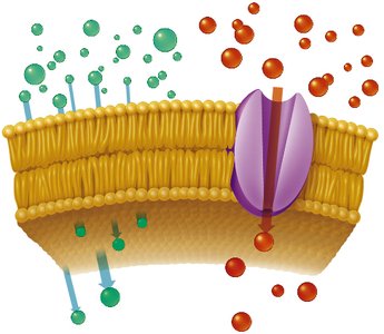
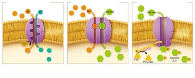
Passive and Active Transport Mechanisms
Cells transport substances across membranes via:
Passive transport: Does not require energy (e.g., simple diffusion, facilitated diffusion)
Active transport: Requires energy to move substances against a concentration gradient
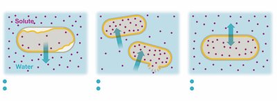
Osmosis
Osmosis is the diffusion of water across a selectively permeable membrane. Water moves from areas of low solute concentration to high solute concentration.
Hypertonic solution: Water leaves the cell, causing plasmolysis.
Hypotonic solution: Water enters the cell, possibly causing lysis if the cell wall is damaged.
Isotonic solution: No net water movement; cell remains unchanged.
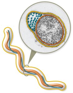
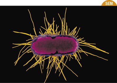
Flagella
Flagella are filamentous structures made of flagellin that provide motility. The basal body anchors the flagellum to the cell wall and plasma membrane. Gram-negative bacteria have a more complex basal body than Gram-positive bacteria.

Flagella Arrangements
Monotrichous: Single flagellum
Lophotrichous: Cluster of flagella at one pole
Amphitrichous: Flagella at both poles
Peritrichous: Flagella all over the cell surface
Periplasmic Flagella (Axial Filaments)
Periplasmic flagella are located between the plasma membrane and cell wall, allowing spirochetes to move in a corkscrew motion.
Fimbriae and Pili
Fimbriae: Short, bristle-like structures for adhesion and biofilm formation; common in Gram-negative bacteria.
Pili: Longer, less numerous structures used for adhesion, movement, and gene transfer (conjugation).
Glycocalyx
The glycocalyx is a sticky, carbohydrate-rich layer outside the cell wall. It can be a loosely associated slime layer or a well-organized capsule. Capsules enhance pathogenicity by protecting cells from phagocytosis and desiccation.
Intracellular Structures of Prokaryotes
Nucleoid
The nucleoid is the region where the prokaryotic chromosome (a single, circular DNA molecule) is located. It is not surrounded by a membrane.
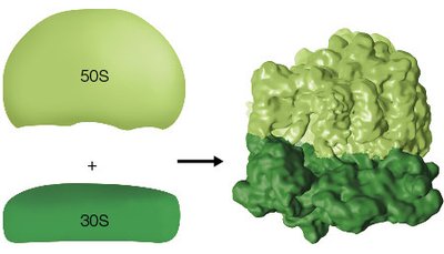
Ribosomes
Prokaryotic ribosomes (70S) are composed of a large (50S) and a small (30S) subunit. They are responsible for protein synthesis and are structurally distinct from eukaryotic ribosomes, supporting the endosymbiotic theory.
Cytoskeleton
The prokaryotic cytoskeleton is made of long protein filaments that provide structural support and help maintain cell shape.
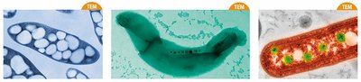
Inclusion Bodies
Inclusion bodies are storage sites for nutrients and other substances. Examples include:
Carboxysomes: Contain enzymes for carbon fixation
Magnetosomes: Contain magnetic iron for orientation in magnetic fields
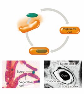
Endospores
Endospores are metabolically inactive, highly resistant structures formed by certain bacteria (e.g., Bacillus anthracis, Clostridium botulinum) in response to stress. They can survive extreme conditions and are a challenge in healthcare settings.
Sporulation: The process of endospore formation, involving DNA replication, packaging, and formation of protective layers.
Germination: When conditions improve, endospores return to vegetative cells.