 Back
BackProkaryotic Cell Structure and Function: Microbiology Study Guide
Study Guide - Smart Notes
 Tailored notes based on your materials, expanded with key definitions, examples, and context.
Prokaryotic vs. Eukaryotic Cells
Overview of Cell Types
Prokaryotic cells and eukaryotic cells differ fundamentally in their structure and organization. Prokaryotes lack a membrane-bound nucleus and organelles, while eukaryotes possess these features. Understanding these differences is essential for microbiology.
Prokaryotic cells: Include bacteria and archaea; DNA is located in a nucleoid region.
Eukaryotic cells: Include plants, animals, fungi, and protists; DNA is enclosed within a nuclear membrane.
Cell envelope: Refers to the layers surrounding the cell, including the cell membrane, cell wall, and glycocalyx.
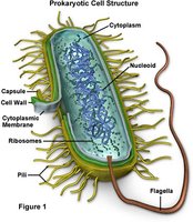
Cell Envelope of Prokaryotic Cells
Cell Membrane
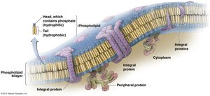
The cell membrane is a critical barrier that separates the interior of the cell from its external environment. In bacteria, it is composed primarily of phospholipids arranged in a bilayer, lacking cholesterol.
Phospholipid bilayer: Provides structural integrity and selective permeability.
Integral and peripheral proteins: Facilitate transport, signaling, and structural support.
Cell Wall
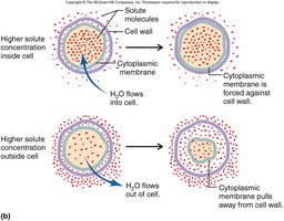
The bacterial cell wall is essential for maintaining cell shape, protecting against osmotic shock, anchoring flagella, affecting pathogenicity, and differentiating between species.
Osmotic protection: Prevents lysis in hypotonic environments and plasmolysis in hypertonic environments.
Species differentiation: Cell wall composition is used in classification (e.g., Gram stain).
Peptidoglycan Structure
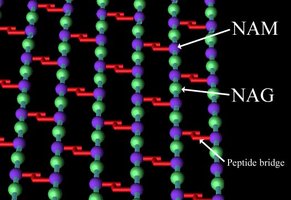
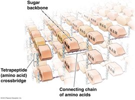
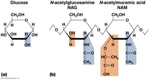
Peptidoglycan is the major component of the bacterial cell wall, consisting of repeating disaccharides: N-acetylglucosamine (NAG) and N-acetylmuramic acid (NAM). These chains are cross-linked by short peptides, providing rigidity.
NAG and NAM: Form the backbone of peptidoglycan.
Peptide bridges: Short chains of amino acids (usually four) connect the sugar chains.
Importance of Peptidoglycan
Peptidoglycan is targeted by antibiotics such as penicillin, which interferes with peptide cross-linking. Differences in cell wall structure are the basis for the Gram stain.
Penicillin: More effective against Gram-positive bacteria due to thick peptidoglycan.
Lysozyme: Enzyme found in saliva, tears, and mucus; breaks NAG-NAM bonds, affecting Gram-positive bacteria.
Gram Positive vs. Gram Negative Cell Walls
Gram Positive Cell Wall
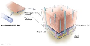
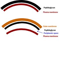
Gram-positive bacteria have a thick peptidoglycan layer and contain teichoic acids, which contribute to cell wall stability and function.
Thick peptidoglycan: Provides structural strength.
Teichoic acids: Play roles in cell wall maintenance and ion regulation.
Gram Negative Cell Wall
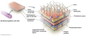
Gram-negative bacteria possess a thin peptidoglycan layer, an outer membrane rich in lipopolysaccharides (LPS), and a periplasmic space. The outer membrane acts as a permeability barrier and protects against many antibiotics.
Outer membrane: Contains LPS, including toxic Lipid A.
Periplasmic space: Region between the outer membrane and cytoplasmic membrane.
Drug resistance: Gram-negative bacteria are less sensitive to many medications.
Other Cell Wall Types
Some bacteria cannot be classified as Gram-positive or Gram-negative. Acid-fast bacteria (e.g., Mycobacterium) have cell walls rich in mycolic acid, making them hydrophobic and difficult to stain. Mycoplasma species lack a cell wall entirely.
Acid-fast cell walls: Contain mycolic acid; resistant to staining and harsh conditions.
No cell wall: Mycoplasma are resistant to antibiotics targeting cell wall synthesis.
Glycocalyx
Structure and Function
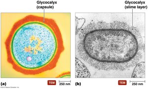
The glycocalyx is a sticky, gelatinous coating surrounding some bacteria. It can be organized as a capsule or a loosely arranged slime layer.
Capsule: Organized, protects against phagocytosis.
Slime layer: Loosely organized, aids in adherence to surfaces.
Cell Attachments
Flagella
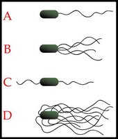
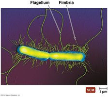
Flagella are motility structures that allow bacteria to move. They can be arranged in various patterns: monotrichous (one), lophotrichous (multiple at one end), amphitrichous (one at each end), and peritrichous (all around).
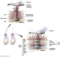
Flagellum structure: Composed of filament, hook, and basal body.
Movement: Requires ATP; allows chemotaxis and phototaxis.
Flagellar antigens: Used for bacterial characterization.
Axial Filaments
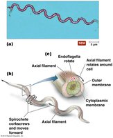
Axial filaments are specialized flagella found in spirochetes, enabling corkscrew motion for movement through viscous environments.
Location: Within the periplasmic space.
Function: Allows snakelike, corkscrew movement.
Fimbriae and Pili
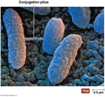
Fimbriae are numerous, short filaments used for attachment, while pili are longer and typically involved in DNA transfer (conjugation).
Fimbriae: Aid in adherence to surfaces and host tissues.
Pili: Facilitate transfer of genetic material between bacteria.
Cytoplasmic Components
DNA and Nucleoid
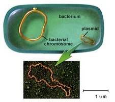
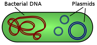
Bacterial DNA is typically a single, circular chromosome located in the nucleoid region. Plasmids are extra-chromosomal DNA elements that often carry genes for antibiotic resistance and toxin production.
Nucleoid: Region containing the chromosome.
Plasmids: Small, circular DNA molecules; can be transferred between cells.
Ribosomes
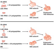
Ribosomes are responsible for protein synthesis. Prokaryotic ribosomes are 70S, while eukaryotic ribosomes are 80S.
70S ribosomes: Composed of 50S and 30S subunits.
Function: Translate mRNA into proteins.
Inclusions
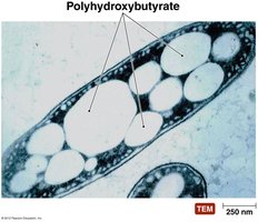
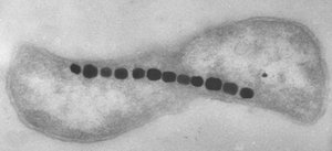
Bacteria may contain various storage granules, such as starch, phosphate, lipid, and sulfur inclusions. These serve as energy reserves or structural materials.
Polyhydroxybutyrate: Lipid storage granule.
Metachromatic granules: Phosphate storage.
Magnetosomes: Magnetic storage for orientation.
Sporulation (Endospore Formation)
Endospore Formation and Function
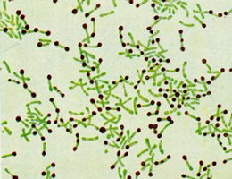
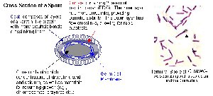
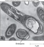
Endospores are dormant, highly resistant forms produced by certain bacteria (e.g., Bacillus and Clostridium). They allow survival under extreme conditions such as nutrient depletion, toxic chemicals, lack of water, and extreme heat.
Resistance: Endospores withstand boiling, desiccation, and chemicals.
Germination: Spores return to vegetative state when conditions improve.
Steps in Sporulation
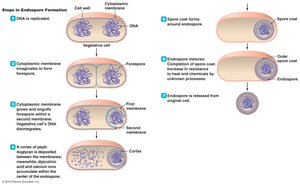
Sporulation involves several steps, including DNA replication, membrane invagination, cortex formation, and spore coat development.
Step 1: DNA is replicated.
Step 2: Membrane grows and forms forespore.
Step 3: Cortex and spore coat form.
Step 4: Mature endospore is released.
