 Back
BackProkaryotic Cell Structure, Antimicrobials, and Bacterial Respiratory Diseases
Study Guide - Smart Notes
 Tailored notes based on your materials, expanded with key definitions, examples, and context.
Introduction to Prokaryotic Cells
Domains of Life
Prokaryotic cells are classified into two domains: Bacteria and Archaea. Eukaryotic cells belong to the domain Eukarya. Prokaryotes are unicellular organisms that lack a membrane-bound nucleus and membrane-bound organelles.
Prokaryotic Cell Structure
Essential and Optional Structures
All bacteria possess certain structures, while others are present only in some species.
Universal structures: Cell membrane, ribosomes, chromosome(s)
Most bacteria: Cell wall
Some bacteria: Flagella, pili, fimbriae, inclusions, endospores
Shapes and Arrangements
Bacteria exhibit three general shapes:
Coccus: Spherical, oval, bean-shaped, or pointed
Bacillus: Cylindrical, filamentous, or club-shaped
Vibrio: Curved
Pleomorphism refers to variations in size and shape among cells of a single species. Mycoplasma species display extreme pleomorphism due to the absence of a cell wall.
Plasma Membrane Structure and Function
Composition and Roles
The plasma membrane is a fluid mosaic composed of a lipid bilayer with embedded proteins. It is selectively permeable and functions in:
Transport (nutrients and waste)
Energy reactions (ATP production)
Nutrient processing and synthesis
Membrane proteins serve as transporters, anchors, receptors, and enzymes.
Cell Wall Structure and Function
Peptidoglycan and Gram Staining
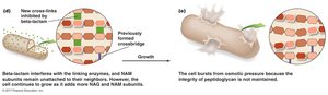
The cell wall determines bacterial shape and provides structural support. Most bacterial cell walls contain peptidoglycan, a unique macromolecule of glycan chains cross-linked by peptides, providing strength and flexibility.
Gram-positive: Thick peptidoglycan layer (20–80 nm), teichoic and lipoteichoic acids, inner cytoplasmic membrane
Gram-negative: Thin peptidoglycan layer (1–3 nm), outer membrane with lipopolysaccharide (LPS), inner cytoplasmic membrane
LPS in Gram-negative bacteria acts as an endotoxin, stimulating fever and shock. Gram-negative bacteria are generally more resistant to chemical agents due to their outer membrane.
Acid-Fast and Atypical Cell Walls
Some bacteria, such as Mycobacterium and Nocardia, have cell walls rich in mycolic acid, making them resistant to chemicals and dyes. Mycoplasma species lack a cell wall and have sterol-enriched membranes, making them resistant to lysis.
Transport Across the Cell Membrane
Passive and Active Transport
Passive transport: No energy required. Includes diffusion (movement from high to low concentration) and osmosis (water movement toward higher solute concentration).
Active transport: Requires energy. Includes primary active transport (uses ATP), secondary active transport (uses ion gradients), and phosphotransferase systems (group translocation).
External Structures for Motility, Adhesion, and Protection
Flagella
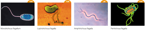
Flagella are protein filaments (flagellin) that provide motility. They consist of a basal body, hook, and filament, and rotate like a propeller. Bacteria move in response to stimuli (chemotaxis, phototaxis, aerotaxis).
Monotrichous: Single flagellum
Lophotrichous: Tufts of flagella
Amphitrichous: Flagella at both poles
Peritrichous: Flagella all over the surface
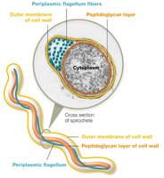
Periplasmic Flagella (Axial Filaments)
Periplasmic flagella are located between the plasma membrane and cell wall, allowing spirochetes to move in a corkscrew motion.
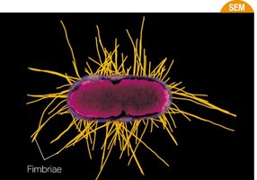
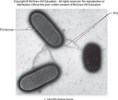
Fimbriae and Pili
Fimbriae are short, bristle-like fibers that aid in adhesion and biofilm formation. Pili are longer, tubular structures used for DNA transfer (conjugation) and attachment.
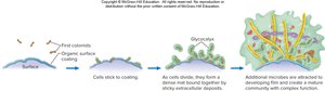
Surface Coatings: Glycocalyx, Slime Layer, and Capsule
The glycocalyx is a sticky, carbohydrate-rich layer aiding in adherence and protection. The slime layer is loosely attached, while the capsule is tightly bound and protects against phagocytosis.
Biofilms
Biofilms are complex communities of microorganisms attached to surfaces and embedded in a self-produced matrix. They protect bacteria from environmental stresses and antibiotics.
Internal Structures of Prokaryotes
Cytoplasm, Chromosome, and Plasmids
The cytoplasm is a gelatinous solution where metabolic activities occur. The bacterial chromosome is typically a single circular DNA molecule in the nucleoid region. Plasmids are small, circular DNA molecules that confer additional traits, such as antibiotic resistance.
Ribosomes
Prokaryotic ribosomes (70S) are composed of a 50S large subunit and a 30S small subunit. They are the site of protein synthesis.
Inclusion Bodies and Cytoskeleton
Inclusion bodies store nutrients and other substances. The cytoskeleton provides structural support and maintains cell shape.
Endospores
Endospores are dormant, highly resistant structures formed by some bacteria (e.g., Bacillus, Clostridium) under stress. They survive extreme conditions and can germinate into vegetative cells when favorable conditions return.
Antibacterial Drugs Targeting the Cell Wall
Mechanisms and Examples
Antibacterial drugs such as penicillins and cephalosporins inhibit peptidoglycan cross-linking, weakening the cell wall and causing lysis. Resistance can occur via beta-lactamases that destroy the beta-lactam ring.
Penicillins: Natural (G, V) and semisynthetic (amoxicillin, ampicillin)
Cephalosporins: Broad spectrum, more resistant to beta-lactamases
Bacitracin, Isoniazid, Vancomycin: Narrow spectrum, used for specific pathogens
Bacterial Respiratory Pathogens with Atypical Cell Walls
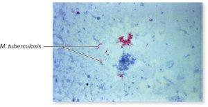
Mycobacterium tuberculosis
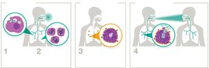
Mycobacterium tuberculosis causes tuberculosis (TB). Its cell wall contains mycolic acids, making it resistant to drying and disinfectants. It can survive inside macrophages by blocking phagolysosome formation.
Clinical Stages of TB
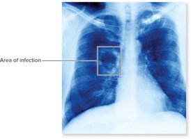
Primary TB: Bacilli are inhaled, phagocytized, and form tubercles (Ghon complexes).
Latent TB: Asymptomatic, non-contagious phase with dormant bacteria.
Active TB: Reactivation leads to severe symptoms and high mortality if untreated.


Diagnosis and Treatment of TB
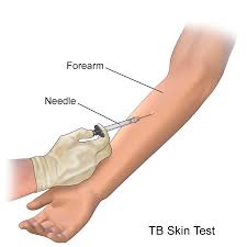
Diagnosis: Skin testing (Mantoux), IGRA, PCR, sputum smears, chest X-rays, Ziehl–Neelsen stain
Treatment: Multi-drug regimens (isoniazid, rifampin, ethambutol, etc.) for up to 9 months
Prevention: Limiting exposure, BCG vaccine (not used in the US)
Mycoplasma pneumoniae
Mycoplasma pneumoniae lacks a cell wall and has a pleomorphic shape. It causes atypical (walking) pneumonia, transmitted by respiratory droplets. Virulence factors include adhesins and hydrogen peroxide production. Diagnosis is by exclusion or serology; treatment is with macrolides if needed.
