 Back
BackProkaryotic Cell Structure: External and Internal Anatomy
Study Guide - Smart Notes
 Tailored notes based on your materials, expanded with key definitions, examples, and context.
Prokaryotic Cell Structure Overview
Introduction
Prokaryotic cells, including bacteria and archaea, possess unique anatomical features that distinguish them from eukaryotic cells. Their structure is essential for survival, adaptation, and pathogenicity. This guide covers the external and internal anatomy of prokaryotic cells, focusing on bacterial cell structure, function, and classification. 
Bacterial Strains and Virulence
Bacterial Strains
A bacterial strain is a genetic variant or subtype within a species, characterized by differences in genetics, metabolism, virulence, or surface proteins. Strains can have significant implications for disease, antibiotic resistance, and beneficial roles in microbiomes.
Commensal vs. Pathogenic Bacteria
Commensal bacteria, such as certain strains of Escherichia coli in the gut, typically do not cause disease in healthy hosts due to the absence of major virulence factors and a non-invasive lifestyle.
Lack of Major Virulence Factors: No toxins, invasion factors, or specialized secretion systems.
Non-Invasive Lifestyle: Remain in gut lumen, do not invade tissues.
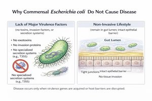 Example: Disease occurs only when virulence genes are acquired or host barriers are disrupted.
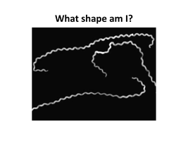
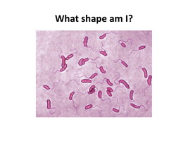
Bacterial Shapes and Arrangements
Major Bacterial Shapes
Bacteria are classified by their shapes:
Bacillus: Rod-shaped
Coccus: Spherical
Spirillus: Spiral-shaped
Bacterial Arrangements
Bacteria may exist singly or in arrangements (chains, clusters) depending on species and division patterns.
Examples of Bacterial Shapes and Arrangements
Bacillus: Rod-shaped bacteria, often found singly or in chains.
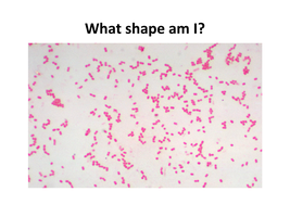
Staphylococcus: Spherical bacteria in grape-like clusters.
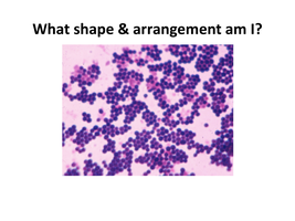
Spirillum: Rigid spiral-shaped bacteria.
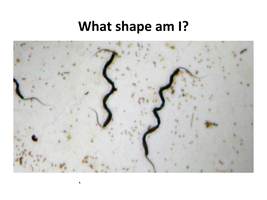
Spirochete: Flexible, tightly coiled spiral bacteria.
Vibrio: Comma-shaped bacteria.
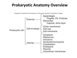
Prokaryotic Anatomy Overview
Structural Components
Prokaryotic cells have both external and internal structures, some of which are present in all bacteria, while others are found only in specific groups.
External: Appendages (flagella, pili, fimbriae), glycocalyx (capsule, slime layer), cell envelope (cell wall, cell membrane)
Internal: Cytoplasm, ribosomes, nucleoid/chromosome, cytoskeleton, endospore, plasmid, intracellular membranes
Cell Membrane Structure and Function
Fluid Mosaic Model
The cell membrane is a dynamic phospholipid bilayer with embedded proteins and other molecules. It regulates transport, supports metabolic processes, and maintains cell integrity.
Phospholipid bilayer forms the basic structure.
Integral & peripheral proteins serve as channels, receptors, and enzymes.
Glycoproteins & glycolipids are involved in cell recognition and signaling.
Cholesterol (in some bacteria) modulates fluidity.
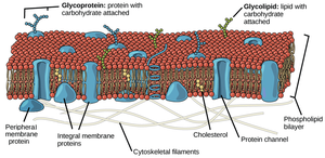 Functions:
Regulates entry and exit of substances
Site for ATP synthesis
Nutrient processing and synthesis
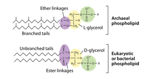
Bacterial vs. Archaeal Cell Membranes
Bacterial Membrane: D-glycerol linked to fatty acids via ester bonds; forms bilayer similar to eukaryotes.
Archaeal Membrane: L-glycerol linked via ether bonds; can form bilayer or monolayer; highly resistant to extreme conditions.
Bacterial Cell Wall and Peptidoglycan
Peptidoglycan Structure
The cell wall provides structural support and protection. Peptidoglycan is a polymer of sugars (NAG and NAM) crosslinked by peptides.
N-acetylglucosamine (NAG) and N-acetylmuramic acid (NAM) form glycan chains.
Peptide bridges crosslink the chains for strength.
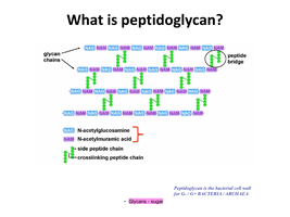 Functions:
Maintains cell shape
Protects against osmotic lysis
Role in cell division
Gram-Positive vs. Gram-Negative Cell Walls
Gram-Positive: Thick peptidoglycan layer, teichoic acids, one membrane, resistant to physical stress.
Gram-Negative: Thin peptidoglycan, outer and inner membranes, lipopolysaccharide (LPS) endotoxin, porins for selective permeability.
Example: Gram-negative bacteria are often more resistant to antibiotics due to the outer membrane and porins.
Summary Table: Bacterial Cell Wall Comparison
Feature | Gram-Positive | Gram-Negative |
|---|---|---|
Peptidoglycan Thickness | Thick | Thin |
Teichoic Acids | Present | Absent |
Outer Membrane | Absent | Present |
LPS (Endotoxin) | Absent | Present |
Porins | Absent | Present |
Additional info:
This guide covers the major external and internal structures of prokaryotic cells, including cell envelope, appendages, and genetic material. It is suitable for exam preparation in a college-level microbiology course, specifically aligned with Chapter 3: Cell Structure and Function.
