 Back
Prokaryotic Cell Structure, Function, and Staining in Microbiology
Study Guide - Smart Notes
 Tailored notes based on your materials, expanded with key definitions, examples, and context.
Prokaryotic Cell Morphology, Structure, and Function
Introduction to Prokaryotes
Prokaryotes are unicellular organisms that lack a membrane-bound nucleus and organelles. They include bacteria and archaea, which are fundamental to the study of microbiology due to their diversity, ecological roles, and medical importance.
Cell Morphology: Prokaryotes exhibit various shapes such as cocci (spherical), bacilli (rod-shaped), spirilla (spiral), vibrios (comma-shaped), and pleomorphic forms.
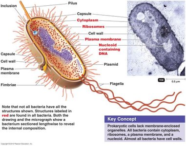
Cell Structure: Key structures include the plasma membrane, cell wall, cytoplasm, nucleoid, ribosomes, and external features like capsules, flagella, fimbriae, and pili.
Function: Each structure contributes to survival, nutrient acquisition, motility, genetic exchange, and pathogenicity.
Microscopy and Cell Size
Relative Sizes of Microbial Cells and Organelles
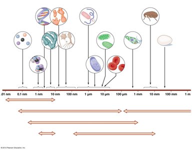
Microorganisms and their components vary greatly in size, requiring different types of microscopy for visualization. Understanding the scale of these structures is essential for selecting appropriate techniques in microbiology.
Microscopy Types: Light microscopes, electron microscopes (SEM, TEM), and atomic force microscopes are used to observe structures from millimeters down to nanometers.
Size Range: Viruses (~0.01–0.3 μm), bacteria and archaea (~0.5–5 μm), eukaryotic cells (10–100 μm), organelles (1–10 μm), and macromolecules (nm scale).
Bacterial Cell Morphology and Arrangements
Common Shapes and Arrangements
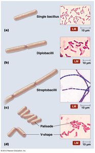
Bacteria display characteristic shapes and arrangements that aid in identification and classification.
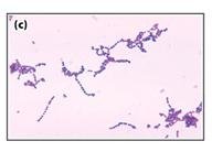
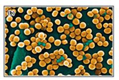
Coccus: Spherical cells; may form chains (streptococci) or clusters (staphylococci).
Bacillus: Rod-shaped cells; may appear singly, in pairs (diplobacilli), or chains (streptobacilli).
Other Forms: Vibrio (comma-shaped), spirillum (rigid spiral), spirochete (flexible spiral), coccobacillus (short rods), and pleomorphic (variable shapes).
Staining Techniques in Microbiology
Purpose and Types of Stains
Staining enhances contrast in microscopic specimens, allowing for visualization of cell morphology, structures, and differentiation between types of bacteria.
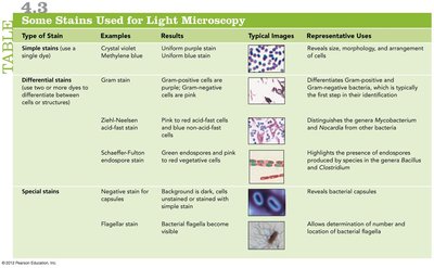
Simple Stains: Use a single dye to reveal cell shape and arrangement.
Differential Stains: Use multiple dyes to distinguish between cell types or structures (e.g., Gram stain, acid-fast stain).
Special Stains: Highlight specific structures such as capsules, flagella, or endospores.
Type of Stain | Examples | Results | Representative Uses |
|---|---|---|---|
Simple stains | Crystal violet, methylene blue | Uniform purple or blue stain | Reveals size, morphology, and arrangement |
Gram stain | Crystal violet, safranin | Gram-positive: purple; Gram-negative: pink | Differentiates Gram-positive and Gram-negative bacteria |
Ziehl-Neelsen acid-fast stain | Carbol fuchsin, methylene blue | Acid-fast: red; Non-acid-fast: blue | Detects Mycobacterium and Nocardia |
Schaeffer-Fulton endospore stain | Malachite green, safranin | Endospores: green; Cells: pink | Highlights endospores in Bacillus and Clostridium |
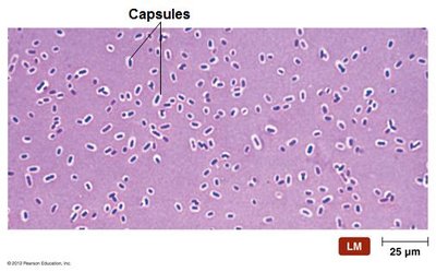
Negative capsule stain | India ink, nigrosin | Capsules appear as clear halos | Reveals bacterial capsules |
Flagella stain | Mordant, carbol fuchsin | Flagella visible | Determines number and arrangement of flagella |
Gram Stain and Cell Wall Structure
Principle and Importance of the Gram Stain
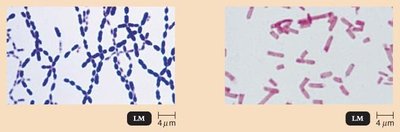
The Gram stain differentiates bacteria based on cell wall structure, which is critical for identification and guiding antimicrobial therapy.
Gram-Positive Bacteria: Thick peptidoglycan layer, retain crystal violet stain (purple).
Gram-Negative Bacteria: Thin peptidoglycan, outer membrane with lipopolysaccharide (LPS), lose crystal violet and take up safranin (pink).
Clinical Relevance: The Gram reaction is one of the first steps in identifying bacterial pathogens in the laboratory.
Bacterial Cell Envelope
Plasma Membrane Structure and Function
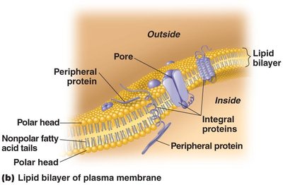
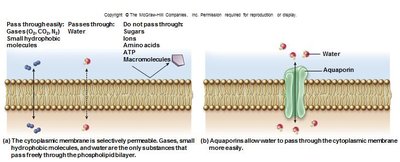
The plasma membrane is a selectively permeable barrier composed of a phospholipid bilayer with embedded proteins. It regulates the movement of substances in and out of the cell and is involved in energy generation and cell signaling.
Fluid Mosaic Model: Describes the dynamic arrangement of lipids and proteins.
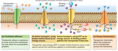
Transport Mechanisms: Include simple diffusion, facilitated diffusion, active transport, and group translocation.
Cell Wall Composition and Function
The bacterial cell wall provides structural support, maintains cell shape, and protects against osmotic lysis. Its composition varies between Gram-positive and Gram-negative bacteria.
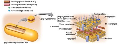
Peptidoglycan: A polymer of N-acetylglucosamine (NAG) and N-acetylmuramic acid (NAM) cross-linked by peptide chains.
Gram-Positive Cell Wall: Thick peptidoglycan with teichoic acids.
Gram-Negative Cell Wall: Thin peptidoglycan, outer membrane with LPS (endotoxin), and periplasmic space.
Specialized Structures of Prokaryotes
Glycocalyx: Capsule and Slime Layer
The glycocalyx is an external layer composed of polysaccharides and/or polypeptides. It can be organized as a capsule or a loose slime layer.
Capsule: Firmly attached, protects against desiccation and phagocytosis, and aids in adherence and biofilm formation.
Slime Layer: Loosely attached, provides similar but less pronounced protection and adherence.
Flagella and Motility
Flagella are long, whip-like appendages that provide motility to bacteria. Their arrangement and structure are important for identification and pathogenicity.
Structure: Composed of filament, hook, and basal body.
Function: Enable movement toward or away from stimuli (taxis), such as chemotaxis, phototaxis, and aerotaxis.
Clinical Relevance: Flagellar proteins can serve as antigens (e.g., H antigen in E. coli O157:H7).
Fimbriae and Pili
Fimbriae and pili are hair-like projections involved in attachment, biofilm formation, motility, and genetic exchange.
Fimbriae: Short, numerous; mediate adherence to surfaces and host tissues.
Pili: Longer, fewer; involved in conjugation (DNA transfer) and sometimes motility.
Internal Structures of Prokaryotes
Nucleoid and Plasmids
The nucleoid is the region containing the bacterial chromosome, while plasmids are small, circular DNA molecules that often carry genes for antibiotic resistance or virulence.
Nucleoid: Not membrane-bound; contains the main genetic material.
Plasmids: Replicate independently; can be transferred between cells via conjugation.
Ribosomes
Bacterial ribosomes (70S) are the sites of protein synthesis, composed of rRNA and proteins. They are targets for several classes of antibiotics.
Storage Granules and Inclusions
Bacteria may store nutrients or building blocks in the form of granules or inclusions, such as glycogen, poly-β-hydroxybutyrate (PHB), phosphate (volutin), and sulfur granules.
Endospores
Endospores are highly resistant, dormant structures formed by some bacteria (e.g., Bacillus and Clostridium) to survive harsh conditions. They are not reproductive but ensure survival through extreme environments.
Structure: Core with DNA, cortex, spore coat, and exosporium; contains calcium dipicolinate for protection.
Formation: Sporulation is triggered by nutrient limitation; germination returns the spore to a vegetative state.
Comparison of Prokaryotic and Eukaryotic Cells
Key Differences
Prokaryotic cells lack membrane-bound organelles and a true nucleus, while eukaryotic cells possess compartmentalized structures for specialized functions.
Prokaryotes: No nucleus, single circular chromosome, 70S ribosomes, cell wall (peptidoglycan in bacteria).
Eukaryotes: True nucleus, multiple linear chromosomes, 80S ribosomes, membrane-bound organelles (mitochondria, ER, Golgi, etc.).
