 Back
BackProkaryotic Cell Surface Structures and Motility Mechanisms
Study Guide - Smart Notes
 Tailored notes based on your materials, expanded with key definitions, examples, and context.
Prokaryotic Cell Surface Structures
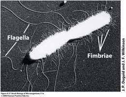
Fimbriae
Fimbriae are short, thin, proteinaceous filaments found on the surface of many bacteria. Their primary function is to facilitate attachment to surfaces, including host tissues and abiotic surfaces, which is crucial for colonization and biofilm formation.
Structure: Composed of protein subunits called pilin.
Function: Mediate adhesion to surfaces and other cells.
Distribution: Numerous and distributed over the entire cell surface.
Example: Escherichia coli uses fimbriae to adhere to the intestinal lining.
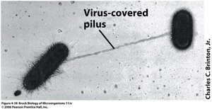
Pili
Pili are longer than fimbriae and are typically present in fewer numbers. They play roles in conjugation (DNA transfer between cells), motility, and adhesion, especially in pathogenic bacteria.
Structure: Composed of pilin proteins; longer and less numerous than fimbriae.
Function: Involved in genetic exchange (conjugation), twitching motility, and adhesion to host tissues.
Type IV Pili: Specialized for motility and DNA uptake.
Example: Neisseria gonorrhoeae uses pili for attachment to human cells.
Unique Attachment Structures in Archaea: Hami
Some Archaea, such as those in the SM1 group, possess unique surface appendages called hami. These are protein filaments similar to pili but end in grappling hook-like structures, facilitating strong attachment within biofilms.
Structure: Filamentous with hook-like ends.
Function: Attachment to surfaces and other cells in biofilms.
Capsules and Slime Layers
Capsules and slime layers are extracellular layers composed of polysaccharides or polypeptides. They provide protection and facilitate attachment to surfaces.
Capsule: A dense, well-organized layer tightly attached to the cell wall; protects against desiccation and phagocytosis.
Slime Layer: A loosely attached, easily removable layer; aids in surface gliding and biofilm formation.
Surface Motility in Prokaryotes
Twitching Motility
Twitching motility is a type of surface movement that requires the extension and retraction of type IV pili. The pili extend from one cell pole, attach to a surface, and retract, pulling the cell forward. This process is powered by ATP hydrolysis.
Mechanism: Extension, attachment, retraction cycle.
Energy Source: ATP hydrolysis.
Examples: Pseudomonas and myxobacteria.
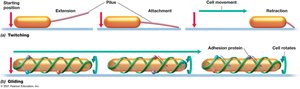
Gliding Motility
Gliding motility is a smooth, continuous movement along the long axis of the cell, independent of external appendages. It is observed only in Bacteria and involves a helical intracellular protein track, gliding motors, and adhesion proteins.
Mechanism: Movement along a protein track with the help of gliding motors and adhesion proteins.
Examples: Myxococcus, Flavobacterium.

Flagella and Prokaryotic Motility
Flagella: Structure and Function
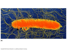
Flagella are long, whip-like appendages used for motility and chemotaxis. Their structure and mechanism of action differ among Bacteria, Archaea, and Eukarya.
Bacterial Flagella: Composed of flagellin; rotate like propellers; powered by proton motive force.
Archaeal Archaella: Structurally similar to type IV pili; powered by ATP; not hollow.
Eukaryotic Flagella and Cilia: Made of microtubules; move by undulation via dynein motor proteins.
Flagellar Arrangements
The arrangement of flagella on the cell surface affects motility patterns:
Monotrichous: Single flagellum at one pole.
Lophotrichous: Cluster of flagella at one or both poles.
Amphitrichous: Single flagellum at both poles.
Peritrichous: Flagella distributed over the entire cell surface.
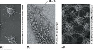
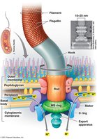
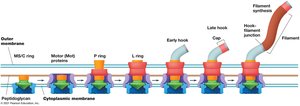
Flagellar Structure and Biosynthesis
Bacterial flagella are complex structures composed of a basal body, hook, and filament. Assembly occurs from the base outward, requiring the coordinated expression of approximately 50 genes.
Basal Body: Anchors the flagellum to the cell envelope and acts as a motor.
Hook: Connects the basal body to the filament.
Filament: Composed of thousands of flagellin subunits.
Energy Source: Proton motive force (Bacteria) or ATP (Archaea).
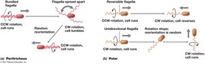
Flagellar Motility Patterns
Flagellar arrangement influences movement patterns:
Polar Flagella: Attached at one or both ends; rapid, spinning movement.
Peritrichous Flagella: Inserted around the cell; slower, straight-line movement.
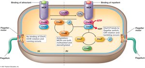
Chemotaxis and Other Taxes
Mechanisms of Chemotaxis
Chemotaxis is the directed movement of motile cells toward or away from chemical stimuli. Cells compare current and past environments to determine movement direction. Other taxes include phototaxis (light), aerotaxis (oxygen), and more.
Chemoreceptors: Detect chemical gradients.
Photoreceptors: Detect light gradients.
Aerotactic Receptors: Detect oxygen gradients.
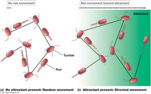
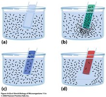
Measuring Chemotaxis
Experimental assays, such as capillary tube assays, are used to measure chemotactic responses by observing bacterial accumulation near attractants or repellents.
Attractant: Bacteria accumulate near the source.
Repellent: Bacteria avoid the source.
Specialized Prokaryotic Structures
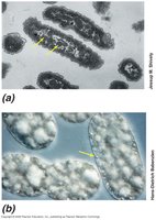
Gas Vesicles
Gas vesicles are protein-bound structures that provide buoyancy to aquatic microorganisms, such as cyanobacteria, allowing them to position themselves optimally for light and nutrients.
Structure: Hollow, rigid, proteinaceous vesicles.
Function: Regulate cell position in the water column.
Magnetosomes
Magnetosomes are intracellular, membrane-bound crystals of magnetic iron minerals that enable magnetotactic bacteria to orient and migrate along Earth's magnetic field lines, typically toward low-oxygen environments.
Structure: Chains of magnetite (Fe3O4) or greigite (Fe3S4) crystals.
Function: Magnetotaxis—orientation and movement along magnetic fields.
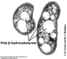
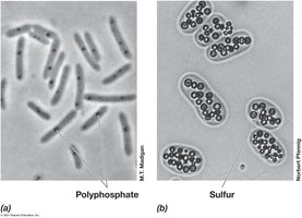
Cell Inclusion Bodies
Inclusion bodies are intracellular storage depots for nutrients and energy reserves, such as glycogen, poly-β-hydroxyalkanoate, polyphosphate, and sulfur granules.
Poly-β-hydroxyalkanoate (PHA): Carbon and energy storage polymer.
Polyphosphate: Phosphate storage.
Sulfur Granules: Store elemental sulfur for energy metabolism.
Carboxysomes
Carboxysomes are protein-based microcompartments found in some autotrophic prokaryotes. They contain the enzyme RuBisCO and are involved in carbon fixation via the Calvin-Benson cycle.
Function: Enhance the efficiency of CO2 fixation.
Example: Found in Halothiobacillus neapolitanus.
Bacterial Endospores
Endospore Formation and Structure
Endospores are highly resistant, dormant structures formed by some Gram-positive bacteria, such as Bacillus and Clostridium, in response to harsh environmental conditions. Sporulation is the process of endospore formation, which occurs within the vegetative cell.
Structure: Contains essential macromolecules, calcium dipicolinic acid, and small acid-soluble proteins; highly dehydrated.
Resistance: Endospores are resistant to heat, chemicals, radiation, and desiccation.
Life Cycle: Vegetative cell → Developing spore → Sporulating cell → Mature spore → Germination (when conditions improve).
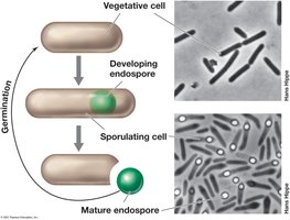
Spore Germination
When environmental conditions become favorable, endospores germinate, returning to the vegetative state. The new vegetative cell is genetically identical to the original cell that produced the spore.
Trigger: Availability of nutrients and suitable conditions.
Process: Spore absorbs water, swells, and resumes metabolic activity.
