Back
BackW7 TH
Study Guide - Smart Notes
 Tailored notes based on your materials, expanded with key definitions, examples, and context.
Protein Catabolism
What are Proteins?
Proteins are large, complex organic molecules composed of amino acids linked by peptide bonds. They are essential for numerous biological functions in all living organisms.
Structural Support: Proteins provide structural integrity to cells and tissues.
Enzymatic Activity: Many proteins function as enzymes, catalyzing biochemical reactions.
Transport: Proteins transport molecules across cell membranes and within the body.
Signaling: Some proteins act as hormones or signaling molecules, regulating physiological processes.
Movement: Motor proteins are involved in muscle contraction and intracellular transport.
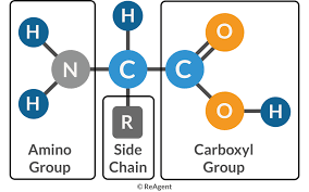
Peptide Bond Formation
Peptide bonds are covalent bonds that link amino acids together in proteins. They are formed through a dehydration synthesis reaction, where the carboxyl group of one amino acid reacts with the amino group of another, releasing a water molecule.
Dehydration Synthesis: Removal of water to form a peptide bond.
Result: Formation of a dipeptide and water.
Equation:
Protein Hydrolysis and Detection in Microbiology
Gelatinase Test
The gelatinase test detects the ability of bacteria to produce gelatinase, an exoenzyme that hydrolyzes gelatin (a protein derived from collagen). This test is important for identifying pathogenic bacteria that can degrade host tissues.
Principle: Gelatinase hydrolyzes gelatin, causing the medium to remain liquid even after refrigeration.
Procedure:
Inoculate nutrient gelatin with the test organism using a stab technique.
Incubate at 37°C for 24–48 hours or longer.
Refrigerate to solidify gelatin in negative controls.
Observe for liquefaction.
Interpretation:
Positive: Medium remains liquid (gelatinase produced).
Negative: Medium solidifies (no gelatinase).
Clinical Relevance: Gelatinase production may help pathogens invade host tissues.
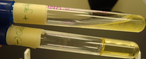
Litmus Milk Test
The litmus milk test is a differential test used to determine how bacteria metabolize lactose, casein, and litmus in milk. It can detect fermentation, proteolysis, reduction, and coagulation reactions.
Components: Skim milk (casein and lactose) and litmus (pH/redox indicator).
Detects:
Lactose fermentation (acid production, pink color).
Casein hydrolysis (peptonization, clearing of medium).
Casein coagulation (clot formation).
Litmus reduction (white color at bottom).
Amino acid catabolism (alkaline, purple color).
Examples:
Pseudomonas aeruginosa: Alkaline reaction (purple).
Lactococcus lactis: Acid reaction (pink).
Streptococcus lactis: Coagulation (clot).
Clostridium sporogenes: Litmus reduction (white).
Bacillus subtilis: Peptonization (clearing).
Urea Hydrolysis and Urease Test
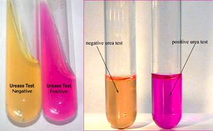
Urease Hydrolysis Test
The urease test identifies bacteria capable of hydrolyzing urea into ammonia and carbon dioxide using the enzyme urease. The production of ammonia raises the pH, which is detected by a color change in the medium.
Reaction:
Indicator: Phenol red (yellow/orange at acidic/neutral pH, pink at alkaline pH > 8.4).
Procedure:
Inoculate urea broth with the test organism.
Incubate at 35–37°C.
Observe color change.
Interpretation:
Positive: Pink/red (urease present, ammonia produced).
Negative: Yellow/orange (no urease activity).
Clinical Relevance: Rapidly distinguishes Proteus (urease positive) from other Enterobacteriaceae; helps identify Helicobacter pylori in gastric ulcers; useful in UTI diagnosis.
Amino Acid Catabolism
Deamination
Deamination is the removal of the amino group from an amino acid, producing ammonia and an organic acid. This process allows bacteria to use amino acids as carbon and energy sources.
Enzyme: Deaminase (endoenzyme).
Products: Ammonia (NH3) and an organic acid.
Significance: Ammonia is excreted; organic acids may be used in metabolism.
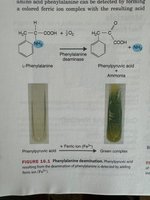
Phenylalanine Deamination
The phenylalanine deaminase test detects the ability of bacteria to deaminate phenylalanine, producing phenylpyruvic acid, which forms a green complex with ferric ions.
Procedure: Inoculate phenylalanine slant, incubate, add ferric chloride.
Positive: Green color (phenylpyruvic acid present).
Negative: No color change.
Examples: Proteus species are positive; most Enterobacteriaceae are negative.
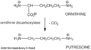
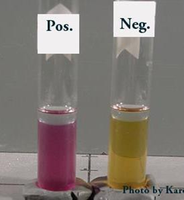
Ornithine Decarboxylation
Decarboxylation is the removal of a carboxyl group (CO2) from an amino acid. Ornithine decarboxylase converts ornithine to putrescine, resulting in an alkaline pH shift detected by a color change in the medium.
Reaction: Ornithine + ornithine decarboxylase → Putrescine + CO2
Indicator: Bromocresol purple (yellow in acid, purple in alkaline).
Procedure: Inoculate medium with glucose, ornithine, and indicator; incubate.
Interpretation:
Positive: Purple (alkaline, putrescine present).
Negative: Yellow (acidic, only glucose fermented).
Examples: E. coli, Enterobacter, Salmonella are positive; Bacillus subtilis, Proteus, Klebsiella, Shigella are negative.
Combined Biochemical Tests
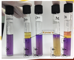
Motility Indole Ornithine (MIO) Test
The MIO test is a single-tube test that simultaneously assesses bacterial motility, indole production, and ornithine decarboxylase activity.
Motility: Diffuse/hazy growth indicates motility; growth only along stab line indicates non-motility.
Indole Production: After incubation, addition of Kovac’s reagent produces a red/pink layer if indole is present.
Ornithine Decarboxylase: Purple color indicates positive decarboxylation; yellow indicates negative.
Examples:
Proteus vulgaris: Motile, Indole positive, Ornithine negative.
Escherichia coli: Motile, Indole positive, Ornithine positive.
Enterobacter aerogenes: Motile, Indole negative, Ornithine positive.