 Back
BackRespiratory and Invasive Bacterial Pathogens: Structure, Pathogenesis, Epidemiology, and Prevention
Study Guide - Smart Notes
 Tailored notes based on your materials, expanded with key definitions, examples, and context.
Introduction to Respiratory and Invasive Bacterial Pathogens
Overview
This module provides a comprehensive overview of major bacterial pathogens affecting the human respiratory system and those capable of causing invasive disease. It covers terminology, host defences, epidemiology, transmission, pathogenesis, clinical outcomes, and prevention strategies, with a focus on Streptococcus pneumoniae, Neisseria meningitidis, Mycobacterium tuberculosis, Haemophilus influenzae, and Bordetella pertussis.
Respiratory System Terminology and Defences
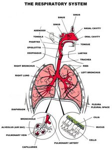
Anatomy of the Respiratory System
The respiratory system is divided into upper and lower tracts, each with specialized structures and defences against microbial invasion.
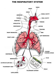
Nasal cavity: Lined with hairs and ciliated mucous membranes to filter, warm, and humidify air, trapping particles and microbes.
Pharynx: Shared pathway for air and food, lined with ciliated mucosa to move contaminants toward the digestive tract.
Tonsils: Lymphoid tissue aggregations that detect and respond to pathogens.
Trachea, bronchi, bronchioles: Conduct air to the lungs; lined with ciliated epithelium and mucus to trap and remove debris.
Alveoli: Tiny air sacs where gas exchange occurs.
Upper Respiratory Tract Defences
Mucus and cilia: Trap and move particles and microbes toward the pharynx for swallowing.
Antimicrobial secretions: Mucus contains lysozyme and defensins that neutralize pathogens.
Resident flora: Includes Haemophilus influenzae and Staphylococcus aureus, which help prevent colonization by harmful microbes.
Lower Respiratory Tract Defences
Mucociliary escalator: Cilia drive mucus upward to the pharynx for removal.
Alveolar macrophages: Phagocytize small particles and pathogens that reach the alveoli.
Risk Factors for Bacterial Respiratory Infections
Viral infections (damage epithelium)
Smoking (impairs cilia)
Immunocompromised state
Age extremes (children, elderly)
Environmental exposure (damp, crowded settings)
Chronic diseases (asthma, diabetes, obesity)
Streptococcus pneumoniae
Biology and Virulence
Gram-positive diplococci
Over 90 serotypes, distinguished by polysaccharide capsule
Capsule is the major virulence factor, protecting against phagocytosis
Antimicrobial resistance is increasing
Clinical Syndromes and Epidemiology
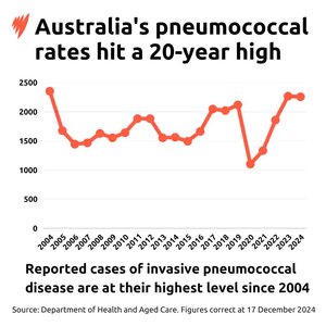
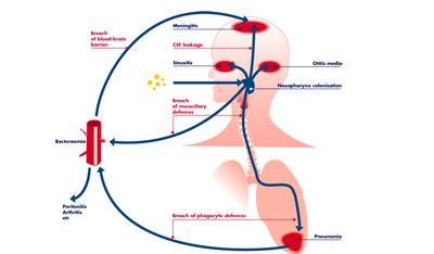
Causes pneumonia, bacteremia, meningitis, and otitis media
Responsible for 30% of community-acquired and up to 50% of hospital-acquired pneumonias
Common complication of influenza and measles
Case fatality rate for invasive disease: 5–7%, higher in elderly
Transmission and Colonization
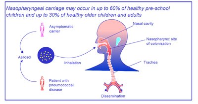
Humans are the only reservoir
Colonizes nasopharynx, especially in children (asymptomatic carriage common)
Spread via respiratory droplets and autoinoculation
More common in winter/early spring
Potential Infection Outcomes
Bacteremia (most common in children)
Meningitis (leading cause in children under 5)
Acute otitis media (major cause in children)
Pneumococcal Disease Outbreaks and Otitis Media
Outbreaks rare, usually in crowded settings
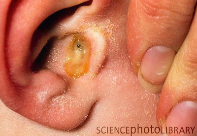
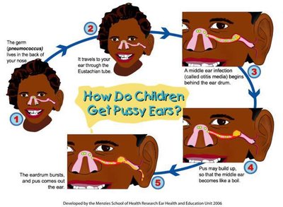
Otitis media: most common reason for antibiotic prescription in children
Linked to nasopharyngeal colonization
Pneumococcal Vaccines
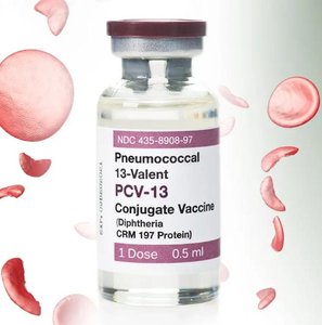
Conjugate vaccines (PCVs) are now standard; polysaccharide vaccines phased out
PCVs induce T-cell dependent immunity and immunologic memory
Highly effective in infants and young children
Included in National Immunisation Programs

Neisseria meningitidis
Biology and Pathogenesis
Gram-negative, non-motile diplococci
Encapsulated; capsule is a major virulence factor
Asymptomatic carriage in nasopharynx is common
Causes invasive disease: meningitis, sepsis, pneumonia, arthritis
Clinical Features and Epidemiology
Rapid progression; can be fatal within hours if untreated
Highest risk: infants, young children, adolescents, elderly
Long-term sequelae in survivors: amputations, brain damage, hearing loss
Sporadic outbreaks, especially in winter/early spring

Pathogenesis
Entry via inhalation of droplets; adheres to non-ciliated epithelial cells using pili and adhesins
Internalized, evades immune detection, crosses mucosal barrier into bloodstream
Capsule protects against phagocytosis and complement
Outer membrane vesicles (OMV) with LOS trigger massive cytokine response, vascular damage, DIC
Serogroups and Vaccines
12 serogroups; A, B, C, W, Y cause >95% of disease
MenB is a leading cause in adolescents/young adults
Vaccines: conjugate for A, C, W, Y; protein-based for B
Epidemiology and Outbreaks
Meningitis Belt in Africa: hyperendemic, periodic epidemics
Serogroup replacement observed after vaccination campaigns
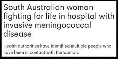
Recent outbreaks in Australia and New Zealand (MenB)
Mycobacterium tuberculosis
Biology and Pathogenesis
Acid-fast, aerobic, non-motile bacilli
Cell wall rich in mycolic acids (waxy, hydrophobic, resistant to disinfectants and antibiotics)
Slow-growing; generation time >20 hours
Transmission and Infection
Spread via inhalation of respiratory droplets
Optimal transmission in crowded, poorly ventilated settings
After inhalation, bacteria reach alveoli, are engulfed by macrophages, and may form granulomas
Latent TB: bacteria dormant, not contagious; risk of reactivation if immunity wanes
Clinical Features and Epidemiology
25% of global population infected (latent TB)
5–10% develop active TB in lifetime
Leading cause of death from a single infectious agent
High burden in Sub-Saharan Africa, Southeast Asia
Treatment and Prevention
Standard regimen: Isoniazid, Rifampicin, Pyrazinamide, Ethambutol (2 months), then Isoniazid and Rifampicin (4 months)
Directly Observed Therapy (DOT) recommended
MDR-TB and XDR-TB require longer, more complex regimens
BCG vaccine: partial protection in children, limited in adults
Haemophilus influenzae
Biology and Pathogenesis
Gram-negative, pleomorphic coccobacillus
Non-motile, facultative anaerobe
Requires X (hemin) and V (NAD+) factors for growth
Capsule (especially type b, Hib) resists phagocytosis
Clinical Features and Epidemiology
Serious infections: meningitis, pneumonia, bacteremia, cellulitis, arthritis
Mild infections: otitis media, sinusitis, conjunctivitis, bronchitis
High-risk: infants <1 year, adults ≥65 years
Prevention and Treatment
Antibiotics for treatment; resistance increasing
Hib conjugate vaccine highly effective; incidence dropped by 99% in children under 5
Non-typeable strains now cause most invasive disease in vaccinated populations
Bordetella pertussis
Biology and Virulence Factors
Gram-negative, pleomorphic coccobacillus
Obligate aerobe, fastidious
Virulence factors: capsule, pertussis toxin (PT), adenylate cyclase toxin (ACT), tracheal cytotoxin (TCT), filamentous hemagglutinin (FHA)
Clinical Features and Epidemiology
Causes pertussis (whooping cough); highly contagious
Serious in infants (<6 months): apnea, respiratory failure, pneumonia, seizures, encephalopathy
Mild in adults/adolescents: prolonged cough
Diagnosis: PCR preferred, culture gold standard, serology in late stages
Prevention and Treatment
Antibiotics: macrolides first-line; resistance rare but emerging
Vaccination: acellular pertussis vaccine (aP) standard; waning immunity a concern
Maternal vaccination protects infants
Booster recommended every 10 years
Summary Table: Key Features of Major Respiratory and Invasive Bacterial Pathogens
Pathogen | Gram Stain & Morphology | Main Diseases | Major Virulence Factor(s) | Prevention |
|---|---|---|---|---|
Streptococcus pneumoniae | Gram-positive diplococci | Pneumonia, meningitis, otitis media, bacteremia | Polysaccharide capsule | Conjugate vaccine (PCV) |
Neisseria meningitidis | Gram-negative diplococci | Meningitis, sepsis, pneumonia | Capsule, LOS, OMV | Conjugate & protein-based vaccines |
Mycobacterium tuberculosis | Acid-fast bacilli | Tuberculosis (pulmonary & extrapulmonary) | Mycolic acid-rich cell wall | BCG vaccine (partial) |
Haemophilus influenzae | Gram-negative coccobacillus | Meningitis, pneumonia, otitis media | Capsule (Hib) | Hib conjugate vaccine |
Bordetella pertussis | Gram-negative coccobacillus | Pertussis (whooping cough) | Pertussis toxin, ACT, TCT | Acellular pertussis vaccine (aP) |
Additional info: This summary integrates and expands upon the provided lecture content, ensuring coverage of all major pathogens, their clinical relevance, and public health implications. Images were included only when directly relevant to the adjacent text and explanation.
