 Back
BackRespiratory System Infections: Microbiology, Pathogenesis, and Clinical Features
Study Guide - Smart Notes
 Tailored notes based on your materials, expanded with key definitions, examples, and context.
Respiratory System Infections
Overview of the Respiratory System
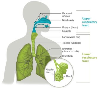
The human respiratory system is divided into upper and lower tracts, each with distinct anatomical features and functions. The respiratory tract is the most common portal of entry for microbes, making it a frequent site of infection. Understanding the anatomy and defense mechanisms of the respiratory system is essential for recognizing how infections develop and spread.
Upper respiratory tract: Includes the paranasal sinuses, nasal cavity, pharynx, and epiglottis. Functions to warm, humidify, and filter air.
Lower respiratory tract: Comprises the larynx, trachea, bronchi, bronchioles, and alveoli. Responsible for directing air to the lungs and facilitating gas exchange.
Factors Limiting Respiratory Infections
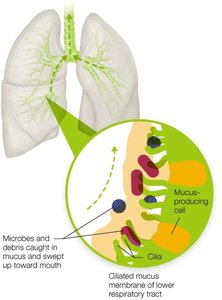
Mucociliary escalator: Ciliated mucous membranes trap and sweep debris and microbes toward the mouth, preventing entry into the lungs.
Alveolar macrophages: Immune cells in the alveoli clear out debris and pathogens not trapped by the mucociliary escalator.
Common Terminology in Respiratory Infections
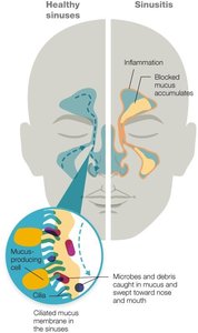
Sinusitis: Inflammation of the sinus membranes, often due to infection or allergens, leading to blocked drainage and mucus accumulation.
Pharyngitis: Inflammation of the pharynx (throat), caused by bacteria, viruses, or allergens.
Epiglottitis: Inflammation and swelling of the epiglottis, which can block the airway and is a medical emergency.
Laryngitis: Inflammation of the larynx, often causing temporary voice loss.
Tracheitis: Inflammation of the trachea.
Bronchitis: Inflammation of the bronchi and/or bronchioles.
Croup: Combined inflammation of the larynx, trachea, bronchi, and bronchioles, usually viral in origin.
Pneumonia: Inflammation of the lung tissue, which can be life-threatening.
Dyspnea: Shortness of breath.
Stridor: Wheezing or loud breathing due to a blocked or narrowed airway.
Respiratory Tract Microbiome
Normal Flora and Its Role
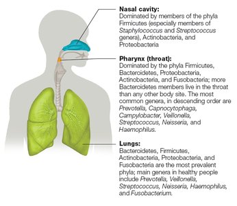
The respiratory tract is colonized by a diverse microbiome, which varies between individuals and health states. The normal flora competes with pathogens and can secrete antimicrobial peptides, providing a protective effect.
Upper respiratory tract: Dominated by Firmicutes, Bacteroidetes, Proteobacteria, Actinobacteria, and Fusobacteria.
Lungs: Once thought sterile, now known to harbor microbiota similar to the mouth.
Viral Infections of the Respiratory System
Common Cold
The common cold is the most frequent acute respiratory infection, caused by over 200 genetically distinct viruses. It is highly infectious and usually self-limiting, but can lead to secondary bacterial infections.
Main causative agents: Rhinoviruses and coronaviruses (60–80% of cases), parainfluenza viruses, adenoviruses, and nonpolio enteroviruses.
Transmission: Personal contact, respiratory droplets, and fomites.
Symptoms: Sore throat, runny nose, cough, sneezing, fatigue, body aches, low-grade fever (especially in children), and thickened mucus in later stages.
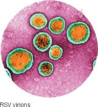
Respiratory Syncytial Virus (RSV)
RSV is a leading cause of acute lower respiratory tract infection in children under 5 and can be severe in infants and the elderly.
Etiological agent: Enveloped RNA virus, Paramyxoviridae family.
Transmission: Respiratory droplets and fomites.
Symptoms: Coughing, sneezing, fever, and wheezing.
Prevention: Sanitary practices, hand washing, and antibody injections for high-risk groups.
Human Parainfluenza Virus (HPIV)
Etiological agent: Single-stranded RNA viruses, Paramyxoviridae family.
Symptoms: Cold-like in adults; croup, bronchitis, and pneumonia in children.
Transmission: Respiratory droplets and fomites.
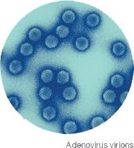
Adenovirus Infections
Etiological agent: Nonenveloped DNA viruses, >50 types infect humans.
Symptoms: Sore throat, cold-like symptoms, conjunctivitis, gastroenteritis, and cystitis.
Prevention: Vaccine for types 4 and 7 (military personnel), sanitary practices.
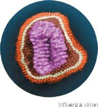
Influenza
Influenza viruses cause seasonal epidemics and occasional pandemics. There are three main types: A, B, and C, with type A being the most significant for human disease.
Virulence factors: Hemagglutinin (HA) and neuraminidase (NA) glycoprotein spikes.
Antigenic drift: Minor mutations in HA and NA, leading to seasonal outbreaks.
Antigenic shift: Major genetic changes, potentially causing pandemics.
Prevention: Annual vaccination (inactivated, recombinant, or live attenuated vaccines).
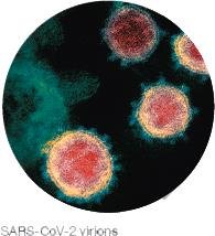
Coronaviruses and COVID-19
Coronaviruses are a large family of enveloped RNA viruses. SARS-CoV-2, the cause of COVID-19, emerged in 2019 and led to a global pandemic.
Transmission: Respiratory droplets and aerosols.
Symptoms: Fever, cough, shortness of breath, fatigue, loss of smell, and more.
Complications: Acute respiratory distress syndrome (ARDS), cytokine storm, multi-organ failure.
Diagnosis: RT-PCR for viral RNA, rapid antigen tests.
Prevention: Vaccination, sanitary practices.
Hantavirus Pulmonary Syndrome (HPS)
Etiological agent: Hantavirus (genus Hantavirus), >40 known viruses.
Transmission: Inhalation of dust containing rodent urine or feces.
Symptoms: Flu-like symptoms, can progress to pulmonary edema and has a high mortality rate (30–40%).

Bacterial Infections of the Respiratory System
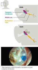
Otitis Media
Otitis media is a common bacterial complication of colds, especially in children due to anatomical differences in the eustachian tube.
Etiological agents: Streptococcus pneumoniae, Moraxella catarrhalis, nontypable Haemophilus influenzae.
Symptoms: Earache, redness, pus or blood leakage, fever.
Prevention: Pneumococcal conjugate vaccine, ear tubes for recurrent infections.
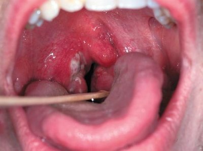
Streptococcus pyogenes and Strep Throat
Etiological agent: Group A Streptococcus (GAS), S. pyogenes.
Symptoms: Sore throat, swollen lymph nodes, fever, exudate in throat/tonsils.
Complications: Scarlet fever, rheumatic fever, otitis, sinusitis, pneumonia.
Diagnosis: Rapid strep test, culture.
Treatment: Penicillin-based or macrolide antibiotics.
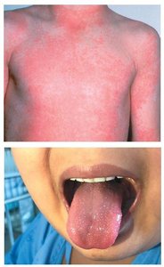
Scarlet Fever
Etiological agent: Lysogenized S. pyogenes producing erythrogenic toxin.
Symptoms: Red sandpaper-like rash, strawberry tongue, rash progression from face/neck down the body.
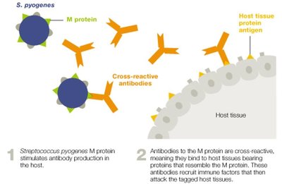
Autoimmune Complications of S. pyogenes
Rheumatic fever: Autoimmune response triggered by cross-reactive antibodies to M protein, affecting joints, heart, and nervous system.
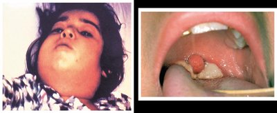
Diphtheria
Etiological agent: Corynebacterium diphtheriae, Gram-positive rod.
Symptoms: Sore throat, low-grade fever, pseudomembrane in airway, "bull neck" appearance.
Complications: A-B exotoxin can cause systemic effects and death if untreated.
Prevention: DTaP vaccine.
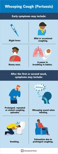
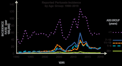
Pertussis (Whooping Cough)
Etiological agent: Bordetella pertussis, Gram-negative bacterium.
Stages: Catarrhal (cold-like), paroxysmal (severe coughing), convalescent (recovery).
Complications: Vomiting, rib fractures, neurological symptoms in children.
Prevention: DTaP/Tdap vaccine, booster recommended for adults and high-risk groups.
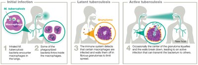
Tuberculosis (TB)
Pathogenesis and Clinical Features
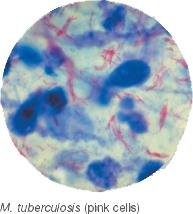
Etiological agent: Mycobacterium tuberculosis, acid-fast rod.
Transmission: Respiratory droplets from active TB patients.
Progression: Most develop latent infection; some progress to active TB with cough, fever, night sweats, weight loss.
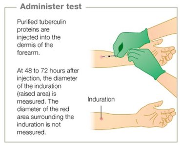
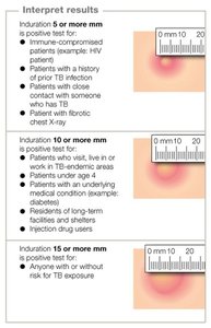
Diagnosis: Mantoux tuberculin skin test, IGRA, molecular diagnostics, chest X-ray, sputum microscopy.
Treatment: Combination antibiotic therapy; MDR and XDR strains require longer, more complex regimens.
Prevention: BCG vaccine in high-risk areas.
Pneumonia
Classification and Clinical Features
Pneumonia is an inflammation of the lower respiratory tract, caused by a variety of microbes. It is classified as typical or atypical based on clinical presentation and causative agents.
Clinical Feature | Typical Pneumonia | Atypical Pneumonia |
|---|---|---|
General Presentation | High fever, sudden onset, productive cough, chest pain | Lower fever, nonproductive cough, subtle onset, muscle aches |
Physical Exam | Respiratory distress, consolidation, crackles | Limited distress, minimal consolidation |
Chest X-ray | Fluid accumulation, opaque areas | Patchy/diffuse pattern, minimal fluid |
Sputum Culture | Often positive for bacteria | Often negative |
Prevalence | 4 out of 5 cases | 1 out of 5 cases |
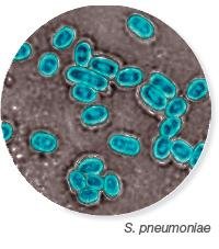
Pneumococcal Pneumonia
Etiological agent: Streptococcus pneumoniae, Gram-positive encapsulated diplococcus.
Prevention: PCV-13 and PPSV-23 vaccines.
Complications: Otitis media, bacteremia, meningitis.
Haemophilus influenzae (Hib) Pneumonia
Etiological agent: Haemophilus influenzae, Gram-negative bacteria.
Prevention: Hib conjugate vaccine.
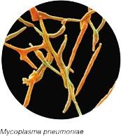
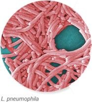
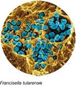
Atypical Bacterial Pneumonia
Common causes: Mycoplasma pneumoniae, Chlamydophila pneumoniae, Legionella pneumophila, Chlamydophila psittaci, Coxiella burnetii, Francisella tularensis.
Transmission: Respiratory droplets, aerosols, zoonotic sources.
Fungal Respiratory System Infections
General Features
Fungal infections (mycoses) of the respiratory tract are rare but increasing due to environmental changes and more immune-compromised individuals. Endemic fungi are geographically restricted, while ubiquitous fungi are found worldwide.
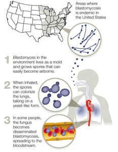
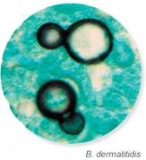
Blastomycosis (Chicago Disease)
Etiological agent: Blastomyces dermatitidis, dimorphic fungus.
Transmission: Inhalation of conidia spores from disturbed soil.
Symptoms: Flu-like, pneumonia-like; can disseminate in immune-compromised patients.
Treatment: Antifungal drugs (amphotericin B, fluconazole, itraconazole, ketoconazole).
Coccidioidomycosis (Valley Fever)
Etiological agent: Coccidioides immitis, C. posadasii.
Transmission: Inhalation of arthroconidia spores from soil.
Symptoms: Fever, cough, joint pain, rash; can disseminate in severe cases.
Treatment: Antifungal drugs for severe cases.
Histoplasmosis
Etiological agent: Histoplasma capsulatum, dimorphic fungus.
Transmission: Inhalation of spores from soil enriched with bat or bird droppings.
Symptoms: Atypical pneumonia-like; most cases asymptomatic.
Treatment: Itraconazole for symptomatic cases.
Aspergillosis
Etiological agent: Aspergillus species, especially A. fumigatus.
Transmission: Inhalation of spores; mainly affects immune-compromised patients.
Symptoms: Fever, cough, hemoptysis, difficulty breathing.
Treatment: Voriconazole, amphotericin B, other antifungals.
Mucormycosis
Etiological agent: Rhizopus arrhizus.
Transmission: Inhalation of spores from rotting wood or soil.
Symptoms: Fever, chest pain, cough, shortness of breath.
Treatment: Amphotericin B.
Pneumocystis Pneumonia (PCP)
Etiological agent: Pneumocystis jirovecii.
Transmission: Likely person-to-person via aerosols.
Symptoms: Fever, fatigue, dyspnea, dry cough; high mortality in untreated cases.
Treatment: Trimethoprim-sulfamethoxazole.
Summary Table: Categorical Classification of SARS-CoV-2 Variants
Category | Examples of Variant’s Attributes | Examples of Public Health Actions |
|---|---|---|
Variants of Interest | Predicted to affect transmission, reduce efficacy of diagnostics/therapeutics, or promote vaccine evasion; limited circulation | Enhanced surveillance, laboratory characterization, continued evaluation |
Variants of Concern | Evidence of reduced efficacy of diagnostics/treatments/vaccines; increased transmissibility or severity | Notify WHO, enhanced testing, research for new vaccines/therapeutics |
Variants of High Consequence | Significant reduction in efficacy of diagnostics/treatments/vaccines; clear increased transmissibility/severity | Notify WHO, announce containment strategies, recommend updates |
Additional info: For all infections, supportive care, early diagnosis, and appropriate antimicrobial or antifungal therapy are critical for optimal outcomes. Vaccination remains a cornerstone for prevention of many respiratory infections.
