 Back
BackRespiratory System Infections: Microbiology, Pathogenesis, and Clinical Features
Study Guide - Smart Notes
 Tailored notes based on your materials, expanded with key definitions, examples, and context.
Respiratory System Infections
Overview of the Respiratory System
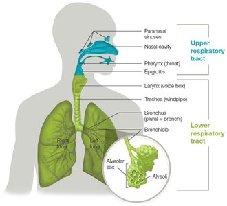
The human respiratory system is divided into upper and lower tracts, each with distinct anatomical structures and functions. The respiratory tract is the most common portal of entry for microbes, making it a frequent site of infection. Understanding the anatomy and defense mechanisms of the respiratory system is essential for recognizing how infections develop and spread.
Upper respiratory tract: Includes the paranasal sinuses, nasal cavity, pharynx, and epiglottis. Functions to warm, humidify, and filter air.
Lower respiratory tract: Comprises the larynx, trachea, bronchi, bronchioles, and alveoli. Responsible for directing air to the lungs and facilitating gas exchange.
Defense Mechanisms of the Respiratory System
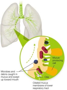
The respiratory system employs several defense mechanisms to limit infection:
Mucociliary escalator: Ciliated mucous membranes trap and sweep debris and microbes toward the mouth for removal.
Alveolar macrophages: Immune cells in the alveoli that engulf and destroy pathogens not trapped by mucus.
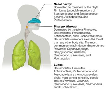
Normal Flora of the Respiratory Tract
The respiratory tract is colonized by a diverse microbiome, which varies between individuals and health states. The normal flora competes with pathogens and can secrete antimicrobial peptides.
Nasal cavity: Dominated by Staphylococcus and Streptococcus species.
Pharynx: Contains Prevotella, Capnocytophaga, Campylobacter, Veillonella, Streptococcus, Neisseria, and Haemophilus.
Lungs: Main genera include Prevotella, Veillonella, Streptococcus, Neisseria, Haemophilus, and Fusobacterium.
Upper Respiratory Tract Infections
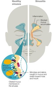
Sinusitis
Sinusitis is inflammation of the sinus membranes, often caused by viral infections or allergens, and less commonly by bacteria. Blocked drainage leads to mucus accumulation and pressure, creating a breeding ground for bacteria.
Symptoms: Congestion, facial pain, pressure, and thick nasal discharge.
Treatment: Antibiotics are only indicated for bacterial cases; most are viral and self-limiting.
Pharyngitis and Epiglottitis
Pharyngitis is inflammation of the pharynx, commonly caused by viruses, bacteria, or allergens. Epiglottitis is a medical emergency involving inflammation of the epiglottis, often due to Haemophilus influenzae type b.
Pharyngitis symptoms: Sore throat, redness, and swelling.
Epiglottitis symptoms: Severe sore throat, difficulty swallowing, and risk of airway obstruction.
Lower Respiratory Tract Infections
Common Terms and Conditions
Laryngitis: Inflammation of the larynx, often causing voice loss.
Tracheitis: Inflammation of the trachea.
Bronchitis: Inflammation of the bronchi and bronchioles.
Croup: Combined inflammation of the larynx, trachea, bronchi, and bronchioles, usually viral in origin.
Pneumonia: Inflammation of lung tissue, potentially life-threatening.
Viral Infections of the Respiratory System
Common Cold
The common cold is an acute respiratory infection caused by over 200 genetically distinct viruses, primarily rhinoviruses and coronaviruses. It is highly infectious and usually self-limiting.
Symptoms: Sore throat, runny nose, cough, sneezing, fatigue, and low-grade fever.
Transmission: Respiratory droplets, personal contact, and fomites.
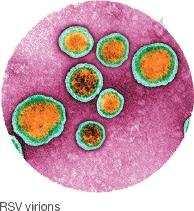
Respiratory Syncytial Virus (RSV)
RSV is an enveloped RNA virus and a leading cause of acute lower respiratory tract infection in children under 5. It can cause severe disease in infants and the elderly.
Symptoms: Coughing, sneezing, fever, and wheezing.
Prevention: Hand hygiene and antibody injections for high-risk groups.
Human Parainfluenza Virus (HPIV)
HPIVs are single-stranded RNA viruses responsible for up to 30% of respiratory infections in children under 5. They can cause croup, bronchitis, and pneumonia.
Transmission: Respiratory droplets and fomites.
Prevention: Supportive care; vaccines are in clinical trials.
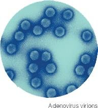
Adenovirus Infections
Adenoviruses are nonenveloped DNA viruses that cause up to 10% of respiratory illnesses in children. They can also cause conjunctivitis, gastroenteritis, and cystitis.
Symptoms: Sore throat, cold-like symptoms, and occasionally severe respiratory disease.
Prevention: Vaccines for military personnel; hygiene practices.
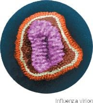
Influenza
Influenza viruses (types A, B, and C) cause seasonal epidemics and occasional pandemics. Type A is the most common cause of epidemics and pandemics, with subtypes defined by hemagglutinin (HA) and neuraminidase (NA) proteins.
Symptoms: Fever, chills, body aches, cough, and fatigue.
Complications: Pneumonia, especially in vulnerable populations.
Prevention: Annual vaccination based on predicted circulating strains.
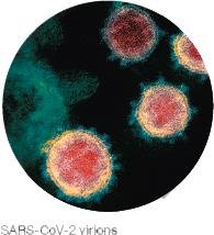
Coronaviruses and COVID-19
Coronaviruses are enveloped RNA viruses. SARS-CoV-2, the cause of COVID-19, emerged in 2019 and is transmitted via respiratory droplets and aerosols. It binds to ACE2 receptors on host cells and can cause severe respiratory distress.
Symptoms: Fever, cough, shortness of breath, fatigue, loss of smell, and gastrointestinal symptoms.
Prevention: Vaccination and hygiene measures.
Hantavirus Pulmonary Syndrome (HPS)
HPS is a rare but severe respiratory illness caused by hantaviruses, transmitted by inhaling dust contaminated with rodent excreta. It can lead to pulmonary edema and has a high mortality rate.
Prevention: Avoiding exposure to rodent habitats and droppings.

Bacterial Infections of the Respiratory System
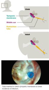
Otitis Media
Otitis media is a common bacterial complication of colds, especially in children due to their shorter and narrower eustachian tubes. It is most often caused by Streptococcus pneumoniae, Moraxella catarrhalis, and nontypable Haemophilus influenzae.
Symptoms: Earache, fever, and sometimes pus or blood leakage.
Prevention: Pneumococcal vaccination and, in recurrent cases, ear tubes for drainage.
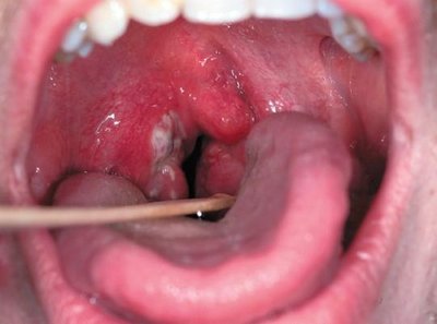
Streptococcus Pyogenes and Strep Throat
Streptococcus pyogenes (Group A Streptococcus) is the main cause of strep throat, which can lead to complications such as otitis, sinusitis, and invasive pneumonia. It is transmitted by respiratory droplets.
Symptoms: Sore throat, swollen lymph nodes, fever, and exudate on tonsils.
Treatment: Penicillin-based antibiotics; macrolides for penicillin-allergic patients.
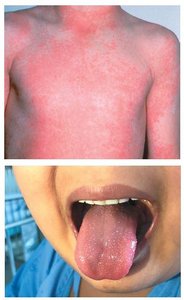
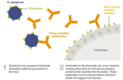
Scarlet Fever and Autoimmune Complications
Scarlet fever is caused by lysogenized S. pyogenes strains producing erythrogenic toxin, leading to a red rash and 'strawberry' tongue. Certain M proteins can trigger autoimmune complications such as rheumatic fever.
Symptoms: Red sandpaper-like rash, strawberry tongue, and peeling skin.
Autoimmunity: Cross-reactive antibodies may attack heart, joints, and kidneys.
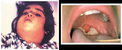
Diphtheria
Diphtheria is caused by Corynebacterium diphtheriae, which produces a pseudomembrane in the upper airway and can release a potent A-B exotoxin. Untreated cases can be fatal.
Symptoms: Sore throat, low-grade fever, 'bull neck,' and pseudomembrane formation.
Prevention: DTaP vaccine.
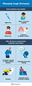
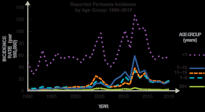
Pertussis (Whooping Cough)
Pertussis is an acute respiratory infection caused by Bordetella pertussis. It progresses through catarrhal, paroxysmal, and convalescent stages, with severe coughing fits that can be life-threatening in infants.
Prevention: DTaP vaccine and Tdap booster for adolescents and adults.
Treatment: Macrolide antibiotics.
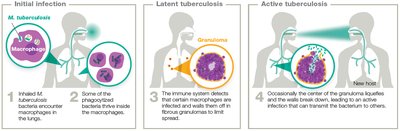
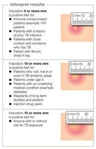
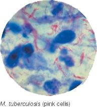
Tuberculosis (TB)
TB is caused by Mycobacterium tuberculosis, an acid-fast rod. It can exist as a latent or active infection, with active TB being highly communicable and potentially fatal. Multidrug-resistant (MDR) and extensively drug-resistant (XDR) strains pose significant treatment challenges.
Diagnosis: Mantoux tuberculin skin test, IGRA, molecular diagnostics, and imaging.
Treatment: Combination antibiotic therapy for several months; longer for resistant strains.
Prevention: BCG vaccine in high-risk regions.

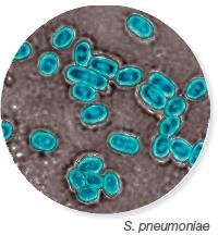
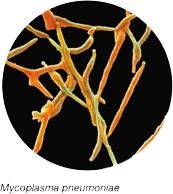
Pneumonia
Pneumonia is inflammation of the alveoli, caused by a variety of microbes. It is classified as typical (with consolidation) or atypical (without consolidation). Community-acquired and healthcare-associated forms exist.
Typical agents: Streptococcus pneumoniae, Haemophilus influenzae.
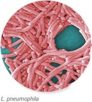
Atypical agents: Mycoplasma pneumoniae, Chlamydophila pneumoniae, Legionella pneumophila, and zoonotic bacteria.
Fungal Respiratory System Infections
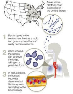
Endemic Mycoses
Endemic fungi cause respiratory infections in specific geographic regions. Examples include:
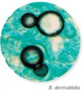
Blastomycosis: Caused by Blastomyces dermatitidis, found in the Mississippi and Ohio River valleys and Great Lakes region.
Coccidioidomycosis (Valley Fever): Caused by Coccidioides immitis and C. posadasii, found in the southwestern U.S. and California.
Histoplasmosis: Caused by Histoplasma capsulatum, found near river valleys and in soil enriched with bat and bird droppings.
Ubiquitous Fungi and Opportunistic Mycoses
Ubiquitous fungi such as Aspergillus and Rhizopus species cause serious infections mainly in immune-compromised patients. Pneumocystis jirovecii causes Pneumocystis pneumonia (PCP), a major concern in HIV/AIDS patients.
Prevention: Avoiding exposure and prophylactic antifungal therapy in high-risk patients.
Summary Table: Clinical Presentation of Pneumonia
Clinical Feature | Typical Pneumonia | Atypical Pneumonia |
|---|---|---|
General Presentation | High fever, sudden onset, productive cough, chest pain, chills | Lower fever, nonproductive cough, subtle onset, runny nose, muscle aches |
Physical Exam | Respiratory distress, consolidation, crackles/rales | Limited distress, minimal consolidation |
Chest X-ray | Consolidation, opaque area in one lobe | Patchy/diffuse pattern, atelectasis, little fluid |
Sputum Culture | Gram-positive or Gram-negative bacteria | Often negative |
Prevalence | 4 out of 5 cases | 1 out of 5 cases |
Additional info: This guide covers the major respiratory system infections, their causative agents, clinical features, and prevention strategies, integrating textbook-level explanations and visual aids for comprehensive exam preparation.
